
Mechanism of tcirg1b deficiency in disrupting central nervous system homeostasis of zebrafish
Received date: 2025-08-28
Revised date: 2025-11-06
Online published: 2025-12-01
Supported by
National Natural Science Foundation of China(31970764)
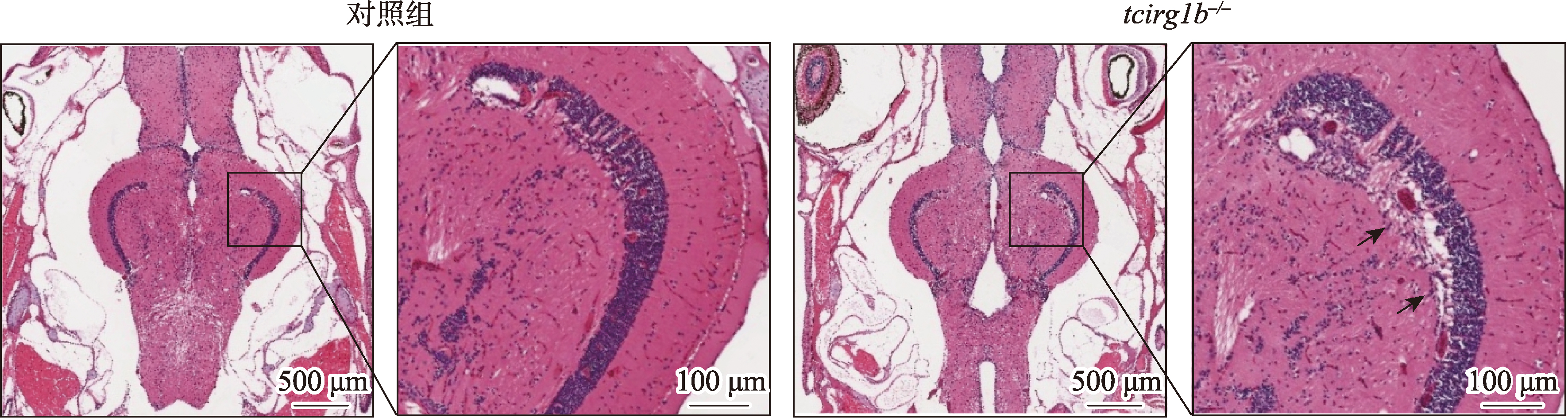
Microglial phagocytosis is crucial for maintaining central nervous system (CNS) homeostasis, a process that depends on normal lysosomal acidification and is precisely regulated by vacuolar-type ATPase (V-ATPase). While mutations in the V-ATPase a3 subunit (encoded by the tcirg1b gene in zebrafish) are a major cause of human malignant osteopetrosis, the subunit’s function in the CNS remains unknown. To investigate the role of the V-ATPase a3 subunit in the zebrafish CNS, we generated a tcirg1b knockout model. Although mutant zebrafish displayed no early neuronal defects, adult brains exhibited significant pathological alterations and behavioral abnormalities. Previous data showed that loss of tcirg1b resulted in enlarged microglia, suggesting potential functional alterations. In this study, transcriptome sequencing analysis of zebrafish macrophages revealed that phagosome formation and intracellular pH regulation pathway genes were significantly down-regulated. Functional analysis confirmed that V-ATPase a3 subunit deficiency impairs lysosomal acidification and digestive function in microglia, leading to the accumulation of apoptotic cell debris and TMR-dextran. Notably, specific restoration of tcirg1b expression in microglia successfully rescued the behavioral phenotypes of mutants, suggesting that the regulation of CNS homeostasis by the V-ATPase a3 subunit is primarily mediated through microglia. In summary, this study provides the first in vivo evidence that tcirg1b deficiency disrupts microglial function, thereby indirectly leading to an imbalance in CNS homeostasis. Our findings reveal a key role for the V-ATPase a3 subunit in regulating neural homeostasis and offer a new theoretical framework for studying the mechanisms of neurological diseases.
Key words: tcirg1b; V-ATPase a3 subunit; microglia; central nervous system disorder; zebrafish
Yujie Li, Feifei Li, Wenqing Zhang, Qi Chen . Mechanism of tcirg1b deficiency in disrupting central nervous system homeostasis of zebrafish[J]. Hereditas(Beijing), 2026 , 48(2) : 201 -212 . DOI: 10.16288/j.yczz.25-234
| [1] | Kuan CY, Roth KA, Flavell RA, Rakic P. Mechanisms of programmed cell death in the developing brain. Trends Neurosci, 2000, 23(7): 291-297. |
| [2] | Ginhoux F, Lim S, Hoeffel G, Low D, Huber T. Origin and differentiation of microglia. Front Cell Neurosci, 2013, 7: 45. |
| [3] | Dos Santos SE, Medeiros M, Porfirio J, Tavares W, Pessôa L, Grinberg L, Leite REP, Ferretti-Rebustini REL, Suemoto CK, Filho WJ, Noctor SC, Sherwood CC, Kaas JH, Manger PR, Herculano-Houzel S. Similar microglial cell densities across brain structures and mammalian species: implications for brain tissue function. J Neurosci, 2020, 40(24): 4622-4643. |
| [4] | Daneman R. The blood-brain barrier in health and disease. Ann Neurol, 2012, 72(5): 648-672. |
| [5] | Dalmau I, Vela JM, González B, Finsen B, Castellano B. Dynamics of microglia in the developing rat brain. J Comp Neurol, 2003, 458(2): 144-157. |
| [6] | Sierra A, Abiega O, Shahraz A, Neumann H. Janus-faced microglia: beneficial and detrimental consequences of microglial phagocytosis. Front Cell Neurosci, 2013, 7: 6. |
| [7] | Herzog C, Pons Garcia L, Keatinge M, Greenald D, Moritz C, Peri F, Herrgen L. Rapid clearance of cellular debris by microglia limits secondary neuronal cell death after brain injury in vivo. Development, 2019, 146(9): dev174698. |
| [8] | Quick JD, Silva C, Wong JH, Lim KL, Reynolds R, Barron AM, Zeng JL, Lo CH. Lysosomal acidification dysfunction in microglia: an emerging pathogenic mechanism of neuroinflammation and neurodegeneration. J Neuroinflammation, 2023, 20(1): 185. |
| [9] | Muench SP, Trinick J, Harrison MA. Structural divergence of the rotary ATPases. Q Rev Biophys, 2011, 44(3): 311-356. |
| [10] | Bowman EJ, O'Neill FJ, Bowman BJ. Mutations of pma-1, the gene encoding the plasma membrane H+-ATPase of Neurospora crassa, suppress inhibition of growth by concanamycin A, a specific inhibitor of vacuolar ATPases. J Biol Chem, 1997, 272(23): 14776-14786. |
| [11] | Chen FQ, Kang R, Liu J, Tang DL. The V-ATPases in cancer and cell death. Cancer Gene Ther, 2022, 29(11): 1529-1541. |
| [12] | Kawasaki-Nishi S, Bowers K, Nishi T, Forgac M, Stevens TH. The amino-terminal domain of the vacuolar proton- translocating ATPase a subunit controls targeting and in vivo dissociation, and the carboxyl-terminal domain affects coupling of proton transport and ATP hydrolysis. J Biol Chem, 2001, 276(50): 47411-47420. |
| [13] | Lupanga U, Röhrich R, Askani J, Hilmer S, Kiefer C, Krebs M, Kanazawa T, Ueda T, Schumacher K. The Arabidopsis V-ATPase is localized to the TGN/EE via a seed plant-specific motif. eLife, 2020, 9: e60568. |
| [14] | Oka T, Toyomura T, Honjo K, Wada Y, Futai M. Four subunit a isoforms of Caenorhabditis elegans vacuolar H+-ATPase. Cell-specific expression during development. J Biol Chem, 2001, 276(35): 33079-33085. |
| [15] | Li YP, Chen W, Liang Y, Li E, Stashenko P. Atp6i- deficient mice exhibit severe osteopetrosis due to loss of osteoclast-mediated extracellular acidification. Nat Genet, 1999, 23(4): 447-451. |
| [16] | Chu A, Zirngibl RA, Manolson MF. The V-ATPase a3 subunit: structure, function and therapeutic potential of an essential biomolecule in osteoclastic bone resorption. Int J Mol Sci, 2021, 22(13): 6934. |
| [17] | Sobacchi C, Schulz A, Coxon FP, Villa A, Helfrich MH. Osteopetrosis: genetics, treatment and new insights into osteoclast function. Nat Rev Endocrinol, 2013, 9(9): 522-536. |
| [18] | Zhang XY, He JW, Fu WZ, Wang C, Zhang ZL. Novel mutations of TCIRG1 cause a malignant and mild phenotype of autosomal recessive osteopetrosis (ARO) in four Chinese families. Acta Pharmacol Sin, 2017, 38(11): 1456-1465. |
| [19] | Lin JH, Yang TS, Zhang WQ, Liu W. Role of different Lyl1 transcripts in zebrafish primitive hematopoiesis. Hereditas(Beijing), 2025, 47(5): 573-588. |
| 林杰豪, 杨童舒, 张文清, 刘伟. Lyl1不同转录本在斑马鱼原始造血中的作用. 遗传, 2025, 47(5): 573-588. | |
| [20] | Thisse C, Thisse B. High-resolution in situ hybridization to whole-mount zebrafish embryos. Nat Protoc, 2008, 3(1): 59-69. |
| [21] | Wu ST, Xue RT, Hassan S, Nguyen TML, Wang TN, Pan HR, Xu J, Liu QF, Zhang WQ, Wen ZL. Il34-Csf1r pathway regulates the migration and colonization of microglial precursors. Dev Cell, 2018, 46(5): 552-563.e4. |
| [22] | Chen Q, Kou HJ, Demy DL, Liu W, Li JC, Wen ZL, Herbomel P, Huang ZB, Zhang WQ, Xu J. The different roles of V-ATPase a subunits in phagocytosis/endocytosis and autophagy. Autophagy, 2024, 20(10): 2297-2313. |
| [23] | Zhang ML, Li XP, Gao LF, Liu J, Bi ZJ, Miao YH, Shan Y, Yu HL. Nobiletin, an activator of the pyruvate kinase isozyme M1/M2 protein, upregulated the glycolytic signalling pathway and alleviated depressive-like behaviour caused by artificial light exposure at night in zebrafish. Food Chem, 2025, 463(Pt 2): 141328. |
| [24] | Raj B, Wagner DE, McKenna A, Pandey S, Klein AM, Shendure J, Gagnon JA, Schier AF. Simultaneous single-cell profiling of lineages and cell types in the vertebrate brain. Nat Biotechnol, 2018, 36(5): 442-450. |
| [25] | Vidal-Itriago A, Radford RAW, Aramideh JA, Maurel C, Scherer NM, Don EK, Lee A, Chung RS, Graeber MB, Morsch M. Microglia morphophysiological diversity and its implications for the CNS. Front Immunol, 2022, 13: 997786. |
| [26] | Westman J, Walpole GFW, Kasper L, Xue BY, Elshafee O, Hube B, Grinstein S. Lysosome fusion maintains phagosome integrity during fungal infection. Cell Host Microbe, 2020, 28(6): 798-812.e6. |
| [27] | Chen YP, Kou YH, Ni Y, Yang HT, Xu CL, Fan HG, Liu HQ. Microglia efferocytosis: an emerging mechanism for the resolution of neuroinflammation in Alzheimer's disease. J Neuroinflammation, 2025, 22(1): 96. |
| [28] | Borst K, Dumas AA, Prinz M. Microglia: immune and non-immune functions. Immunity, 2021, 54(10): 2194-2208. |
| [29] | Guzmán-Ruíz MA, Guerrero Vargas NN, Ramírez-Carreto RJ, González-Orozco JC, Torres-Hernández BA, Valle- Rodríguez M, Guevara-Guzmán R, Chavarría A. Microglia in physiological conditions and the importance of understanding their homeostatic functions in the arcuate nucleus. Front Immunol, 2024, 15: 1392077. |
| [30] | Yin J, Valin KL, Dixon ML, Leavenworth JW. The role of microglia and macrophages in CNS homeostasis, autoimmunity, and cancer. J Immunol Res, 2017, 5150678. |
| [31] | Serra EL, Medalha CC, Mattioli R. Natural preference of zebrafish (Danio rerio) for a dark environment. Braz J Med Biol Res, 1999, 32(12): 1551-1553. |
| [32] | Scimeca JC, Franchi A, Trojani C, Parrinello H, Grosgeorge J, Robert C, Jaillon O, Poirier C, Gaudray P, Carle GF. The gene encoding the mouse homologue of the human osteoclast-specific 116-kDa V-ATPase subunit bears a deletion in osteosclerotic (oc/oc) mutants. Bone, 2000, 26(3): 207-213. |
| [33] | Falace A, Volpedo G, Scala M, Zara F, Striano P, Fassio A. V-ATPase dysfunction in the brain: genetic insights and therapeutic opportunities. Cells, 2024, 13(17): 1441. |
| [34] | Aoto K, Kato M, Akita T, Nakashima M, Mutoh H, Akasaka N, Tohyama J, Nomura Y, Hoshino K, Ago Y, Tanaka R, Epstein O, Ben-Haim R, Heyman E, Miyazaki T, Belal H, Takabayashi S, Ohba C, Takata A, Mizuguchi T, Miyatake S, Miyake N, Fukuda A, Matsumoto N, Saitsu H. ATP6V0A1 encoding the a1-subunit of the V0 domain of vacuolar H+-ATPases is essential for brain development in humans and mice. Nat Commun, 2021, 12(1): 2107. |
| [35] | Bagh MB, Peng SY, Chandra G, Zhang ZJ, Singh SP, Pattabiraman N, Liu AY, Mukherjee AB. Misrouting of v-ATPase subunit V0a1 dysregulates lysosomal acidification in a neurodegenerative lysosomal storage disease model. Nat Commun, 2017, 8(1): 14612. |
| [36] | Koçak G, Güzel BN, Mıhçı E, Küpesiz OA, Yalçın K, Manguoğlu AE. TCIRG1 and SNX10 gene mutations in the patients with autosomal recessive osteopetrosis. Gene, 2019, 702: 83-88. |
| [37] | Goldmann T, Wieghofer P, Jordão MJC, Prutek F, Hagemeyer N, Frenzel K, Amann L, Staszewski O, Kierdorf K, Krueger M, Locatelli G, Hochgerner H, Zeiser R, Epelman S, Geissmann F, Priller J, Rossi FMV, Bechmann I, Kerschensteiner M, Linnarsson S, Jung S, Prinz M. Origin, fate and dynamics of macrophages at central nervous system interfaces. Nat Immunol, 2016, 17(7): 797-805. |
| [38] | Gao C, Jiang JW, Tan YY, Chen SD. Microglia in neurodegenerative diseases: mechanism and potential therapeutic targets. Signal Transduct Target Ther, 2023, 8(1): 359. |
| [39] | Brockerhoff SE, Dowling JE, Hurley JB. Zebrafish retinal mutants. Vision Res, 1998, 38(10): 1335-1339. |
| [40] | Gestri G, Link BA, Neuhauss SCF. The visual system of zebrafish and its use to model human ocular diseases. Dev Neurobiol, 2012, 72(3): 302-327. |
| [41] | Martin A, Babbitt A, Pickens AG, Pickett BE, Hill JT, Suli A. Single-cell RNA sequencing characterizes the molecular heterogeneity of the larval zebrafish optic tectum. Front Mol Neurosci, 2022, 15: 818007. |
| [42] | Tesmer AL, Fields NP, Robles E. Input from torus longitudinalis drives binocularity and spatial summation in zebrafish optic tectum. BMC Biol, 2022, 20(1): 24. |
| [43] | Walton ZE, Brooks RC, Dang CV. mTOR senses intracellular pH through lysosome dispersion from RHEB. Bioessays, 2019, 41(7): e1800265. |
| [44] | Yambire KF, Rostosky C, Watanabe T, Pacheu-Grau D, Torres-Odio S, Sanchez-Guerrero A, Senderovich O, Meyron-Holtz EG, Milosevic I, Frahm J, West AP, Raimundo N. Impaired lysosomal acidification triggers iron deficiency and inflammation in vivo. eLife, 2019, 8: e51031. |
| [45] | Zhang WJ, Bai JW, Hang K, Xu JX, Zhou CW, Li LJ, Wang ZX, Wang YB, Wang KB, Xue DT. Role of lysosomal acidification dysfunction in mesenchymal stem cell senescence. Front Cell Dev Biol, 2022, 10: 817877. |
/
| 〈 |
|
〉 |

www.chinagene.cn
备案号:京ICP备09063187号
