

一例智力障碍和语言发育迟缓患儿的STAG1基因变异分析
收稿日期: 2025-05-19
修回日期: 2025-07-08
网络出版日期: 2025-07-30
Analysis of STAG1 gene variants identified in a patient with intellectual disability and speech delay
Received date: 2025-05-19
Revised date: 2025-07-08
Online published: 2025-07-30
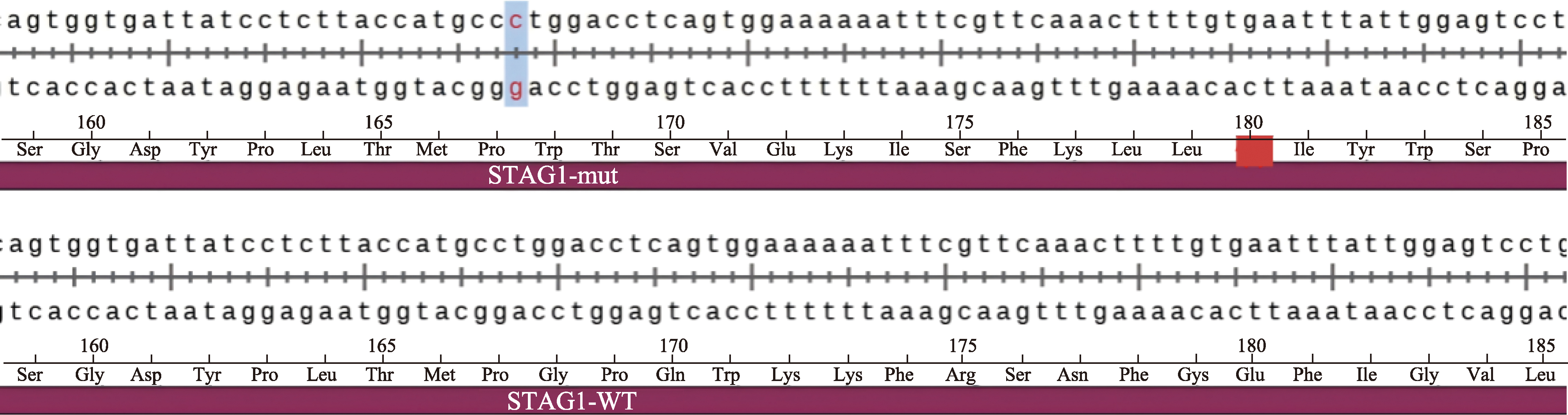
显性遗传智力发育障碍47型(OMIM #617635)与STAG1基因(OMIM*604358)的杂合变异相关,主要临床表现为智力低下、语言发育迟缓、面容异常、癫痫等。本研究报道了1例被诊断为“精神运动发育迟缓”的3岁患儿。利用全外显子组测序技术结合Sanger测序进行基因检测分析发现,该患儿携带STAG1基因c.500dup (p.Gly168TrpfsTer13)为新发变异(其父母均未携带该变异)。根据美国医学遗传学与基因组学学会的遗传变异分类标准与指南,该变异被判定为致病变异(PVS1+PM2_Supporting+PS2_Supporting)。同时,本研究进一步构建突变型质粒表达载体,转染HEK293T细胞,初步发现该变异会导致突变型mRNA表达显著降低,正常STAG1蛋白无法产生。上述研究结果表明,STAG1基因变异导致的临床表型多样,STAG1基因变异的致病机制可能与基因单倍剂量不足相关。
姜翠翠, 吴轲 . 一例智力障碍和语言发育迟缓患儿的STAG1基因变异分析[J]. 遗传, 2026 , 48(2) : 227 -235 . DOI: 10.16288/j.yczz.25-130
Autosomal dominant intellectual developmental disorder (type 47) (OMIM #617635) is associated with heterozygous variants in the STAG1 gene (OMIM*604358). The main clinical manifestations include intellectual disability, delayed language development, abnormal facial features, epilepsy, etc. In this study, we performed the whole-exome sequencing (WES) on a 3-year-old child diagnosed with psychomotor developmental delay, and verified candidate gene variants by Sanger sequencing. We constructed a mutant plasmid expression vector and subsequently transfected it into HEK293T cells to preliminarily investigate the pathogenic mechanism of the identified variant. Additionally, we conducted a comprehensive review of previously published literature to systematically summarize the associated clinical phenotypes. WES revealed that the patient harbored the c.500dup (p.Gly168TrpfsTer13) of the STAG1 gene, and it was a de novo variant. Based on the guidelines from the American College of Medical Genetics and Genomics (ACMG), the variant was classified as a pathogenic variant (PVS1+PM2_Supporting+PS2_Supporting). In vitro cellular function studies have demonstrated that this variant resulted in a substantial decrease in the expression of mutant mRNA, thereby impairing the production of functional STAG1 protein. Clinical phenotypes associated with STAG1 gene variants display considerable variability. This study suggests that the pathogenic mechanism of STAG1 gene variants may be linked to haploinsufficiency of the gene.
| [1] | Lehalle D, Mosca-Boidron AL, Begtrup A, Boute- Benejean O, Charles P, Cho MT, Clarkson A, Devinsky O, Duffourd Y, Duplomb-Jego L, Gérard B, Jacquette A, Kuentz P, Masurel-Paulet A, McDougall C, Moutton S, Olivié H, Park SM, Rauch A, Revencu N, Rivière JB, Rubin K, Simonic I, Shears DJ, Smol T, Taylor Tavares AL, Terhal P, Thevenon J, Van Gassen K, Vincent-Delorme C, Willemsen MH, Wilson GN, Zackai E, Zweier C, Callier P, Thauvin-Robinet C, Faivre L. STAG1mutations cause a novel cohesinopathy characterised by unspecific syndromic intellectual disability. J Med Genet, 2017, 54(7): 479-488. |
| [2] | Li H, Durbin R. Fast and accurate short read alignment with Burrows-Wheeler transform. Bioinformatics, 2009, 25(14): 1754-1760. |
| [3] | McKenna A, Hanna M, Banks E, Sivachenko A, Cibulskis K, Kernytsky A, Garimella K, Altshuler D, Gabriel S, Daly M, DePristo MA. The Genome Analysis Toolkit: a MapReduce framework for analyzing next-generation DNA sequencing data. Genome Res, 2010, 20(9): 1297-1303. |
| [4] | Kadalayil L, Rafiq S, Rose-Zerilli MJJ, Pengelly RJ, Parker H, Oscier D, Strefford JC, Tapper WJ, Gibson J, Ennis S, Collins A. Exome sequence read depth methods for identifying copy number changes. Brief Bioinform, 2015, 16(3): 380-392. |
| [5] | Wang K, Li MY, Hakonarson H. ANNOVAR: functional annotation of genetic variants from high-throughput sequencing data. Nucleic Acids Res, 2010, 38(16): e164. |
| [6] | Richards S, Aziz N, Bale S, Bick D, Das S, Gastier-Foster J, Grody WW, Hegde M, Lyon E, Spector E, Voelkerding K, Rehm HL, ACMG Laboratory Quality Assurance Committee. Standards and guidelines for the interpretation of sequence variants: a joint consensus recommendation of the American College of Medical Genetics and Genomics and the Association for Molecular Pathology. Genet Med, 2015, 17(5): 405-424. |
| [7] | Carramolino L, Lee BC, Zaballos A, Peled A, Barthelemy I, Shav-Tal Y, Prieto I, Carmi P, Gothelf Y, González de Buitrago G, Aracil M, Márquez G, Barbero JL, Zipori D. SA-1, a nuclear protein encoded by one member of a novel gene family: molecular cloning and detection in hemopoietic organs. Gene, 1997, 195(2): 151-159. |
| [8] | Piché J, Van Vliet PP, Pucéat M, Andelfinger G. The expanding phenotypes of cohesinopathies: one ring to rule them all! Cell Cycle, 2019, 18(21): 2828-2848. |
| [9] | Samejima K, Gibcus JH, Abraham S, Cisneros-Soberanis F, Samejima I, Beckett AJ, Pučeková N, Abad MA, Spanos C, Medina-Pritchard B, Paulson JR, Xie LF, Jeyaprakash AA, Prior IA, Mirny LA, Dekker J, Goloborodko A, Earnshaw WC. Rules of engagement for condensins and cohesins guide mitotic chromosome formation. Science, 2025, 388(6743): eadq1709. |
| [10] | Nagano M, Hansen AS. Distance matters: how protein regulators facilitate enhancer-promoter interactions and transcription. Cell Genom, 2025, 5(3): 100817. |
| [11] | Guérin TM, Uhlmann F. Cohesinopathies: is enhancer disruption to blame? Curr Biol, 2025, 35(6): R214-R216. |
| [12] | Yuan B, Neira J, Pehlivan D, Santiago-Sim T, Song XF, Rosenfeld J, Posey JE, Patel V, Jin WH, Adam MP, Baple EL, Dean J, Fong CT, Hickey SE, Hudgins L, Leon E, Madan-Khetarpal S, Rawlins L, Rustad CF, Stray- Pedersen A, Tveten K, Wenger O, Diaz J, Jenkins L, Martin L, McGuire M, Pietryga M, Ramsdell L, Slattery L, DDD Study, Abid F, Bertuch AA, Grange D, Immken L, Schaaf CP, Van Esch H, Bi WM, Cheung SW, Breman AM, Smith JL, Shaw C, Crosby AH, Eng C, Yang YP, Lupski JR, Xiao R, Liu PF. Clinical exome sequencing reveals locus heterogeneity and phenotypic variability of cohesinopathies. Genet Med, 2019, 21(3): 663-675. |
| [13] | Di Muro E, Palumbo P, Benvenuto M, Accadia M, Di Giacomo MC, Manieri S, Abate R, Tagliente M, Castellana S, Mazza T, Carella M, Palumbo O. Novel STAG1 frameshift mutation in a patient affected by a syndromic form of neurodevelopmental disorder. Genes (Basel), 2021, 12(8): 1116. |
| [14] | Funato M, Uehara T, Okada Y, Kaneko H, Kosaki K. Cohesinopathy presenting with microtia, facial palsy, and hearing loss caused by STAG1pathogenic variant. Congenit Anom (Kyoto), 2022, 62(2): 82-83. |
| [15] | Cipriano L, Russo R, Andolfo I, Manno M, Piscopo R, Iolascon A, Piscopo C. A novel de novo STAG1variant in monozygotic twins with neurodevelopmental disorder: new insights in clinical heterogeneity. Genes (Basel), 2024, 15(9): 1184. |
| [16] | Bregvadze K, Sukhiashvili A, Lartsuliani M, Melikidze E, Tkemaladze T. A novel STAG1variant associated with congenital clubfoot and microphthalmia:a case report. SAGE Open Med Case Rep, 2024, 12: 2050313X241277123. |
| [17] | Seymour H, Feben C, Nevondwe P, Kerr R, Spencer C, Mudau M, Honey E, Lombard Z, Krause A, Carstens N. Mutation profiling in south african patients with cornelia de lange syndrome phenotype. Mol Genet Genomic Med, 2024, 12(1): e2342. |
| [18] | Remeseiro S, Cuadrado A, Carretero M, Martínez P, Drosopoulos WC, Cañamero M, Schildkraut CL, Blasco MA, Losada A. Cohesin-SA1 deficiency drives aneuploidy and tumourigenesis in mice due to impaired replication of telomeres. EMBO J, 2012, 31(9): 2076-2089. |
| [19] | Cheng MM, Cao YY. The NMD escape mechanism and its application in disease therapy. Hereditas(Beijing), 2020, 42(4): 354-362. |
| 程苗苗, 曹延延. NMD逃逸机制及其在疾病治疗中的应用. 遗传, 2020, 42(4): 354-362. | |
| [20] | Kurosaki T, Maquat LE. Nonsense-mediated mRNA decay in humans at a glance. J Cell Sci, 2016, 129(3): 461-467. |
/
| 〈 |
|
〉 |

www.chinagene.cn
备案号:京ICP备09063187号-4
总访问:,今日访问:,当前在线:
