

基于斑马鱼模型探究synpo在颅内出血中的作用机制
收稿日期: 2025-10-24
修回日期: 2025-12-02
网络出版日期: 2026-01-30
基金资助
国家自然科学基金项目(32470889);广东省本科高校教学质量与教学改革工程建设项目(粤教高函[2024]9号);广东省本科高校教学质量与教学改革工程建设项目(粤教高函[2024]30号);广东医科大学本科生创新创业教育基地项目(2JD25178);广东医科大学附属医院高层次人才启动项目(2081Z20230049)
The mechanism of synpo in intracerebral hemorrhage using a zebrafish model
Received date: 2025-10-24
Revised date: 2025-12-02
Online published: 2026-01-30
Supported by
National Natural Science Foundation of China(32470889);Project of Teaching Quality and Teaching Reform Engineering for Undergraduate Universities in Guangdong Province (Yue Jiao Gao Han [2024] No. 9);Project of Teaching Quality and Teaching Reform Engineering for Undergraduate Universities in Guangdong Province (Yue Jiao Gao Han [2024] No. 30);Undergraduate Innovation and Entrepreneurship Education Base Project of Guangdong Medical University(2JD25178);High-Level Talents Scientific Research Start-Up Funds of the Affiliated Hospital of Guangdong Medical University(2081Z20230049)
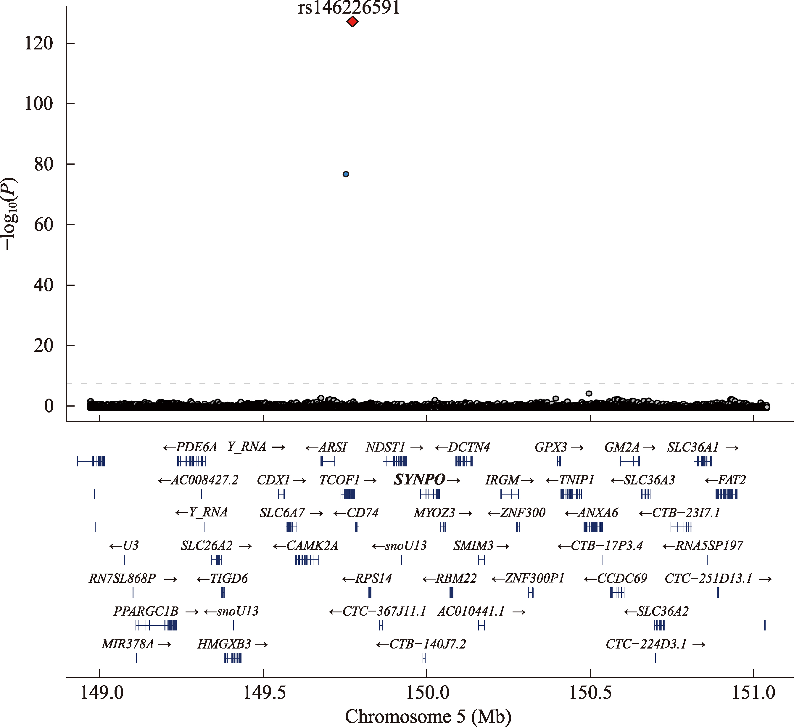
颅内出血(intracerebral hemorrhage,ICH)是一种高致死率的卒中类型,其核心病理机制涉及脑血管稳态受损。遗传因素在ICH疾病发生中占据重要地位,因此挖掘关键调控因子、完善ICH的病理机制网络尤为重要。本研究通过全基因组关联分析,筛选到与ICH显著相关的遗传易感基因——突触足蛋白(synaptopodin)编码基因SYNPO。既往研究表明,SYNPO作为一种进化保守的肌动蛋白结合蛋白,在脑血管内皮细胞中高表达,通过调控肌动蛋白骨架维持内皮细胞连接稳态。然而,其在ICH中的具体作用尚不明确。为此,本研究进一步利用CRISPR/Cas9技术构建了synpo突变体斑马鱼。在肾上腺素刺激下,synpo突变体幼鱼脑血管渗漏率和成鱼ICH发生率都显著高于野生型。转录组分析发现,synpo突变体脑内细胞黏附通路关键基因cdh2显著下调,回补cdh2 mRNA可有效挽救突变体幼鱼的脑血管渗漏。本研究揭示了synpo通过正向调控cdh2维持脑血管稳态,明确“synpo-cdh2”轴是ICH的关键调控通路,为解析ICH的遗传机制及发现潜在干预靶点提供新视角。
欧阳沛东, 唐嘉兰, 吴成超, 李晓玉, 柯珊珊, 张晶晶 . 基于斑马鱼模型探究synpo在颅内出血中的作用机制[J]. 遗传, 2026 , 48(4) : 407 -420 . DOI: 10.16288/j.yczz.25-281
Intracerebral Hemorrhage (ICH) is a stroke subtype with high mortality, and its core pathological mechanism involves the disruption of cerebrovascular homeostasis. Genetic factors play a crucial role in ICH pathogenesis, underscoring the importance of identifying core regulatory factors and delineating the associated pathological network. Here, through genome-wide association study (GWAS), we identified synaptopodin (SYNPO) as a genetic susceptibility gene for ICH. SYNPO is an evolutionarily conserved actin-binding protein previously shown to be highly expressed in cerebrovascular endothelial cells, where it regulates the actin cytoskeleton to maintain endothelial junction stability. However, its functional role in ICH remains unclear. To investigate this, we conducted a synpo mutant zebrafish line using CRISPR/Cas9. Following epinephrine challenge, synpo mutant larvae displayed significantly elevated cerebrovascular leakage compared with wild-type controls, and adult mutants showed a markedly higher incidence of ICH. Transcriptomic profiling revealed significant downregulation of the key adhesion gene cdh2 in mutant brains. Subsequent rescue experiments confirmed that cdh2 mRNA supplementation effectively ameliorated the cerebrovascular leakage. In summary, our study unveils a pathway in which synpo maintains cerebrovascular homeostasis by positively regulating cdh2, demonstrating that the synpo-cdh2 axis serves as a key regulatory pathway in ICH. These findings provide insights into the genetic mechanisms underlying ICH and highlight potential therapeutic targets.
Key words: intracerebral hemorrhage; synpo; cdh2; zebrafish model
| [1] | Puy L, Parry-Jones AR, Sandset EC, Dowlatshahi D, Ziai W, Cordonnier C. Intracerebral haemorrhage. Nat Rev Dis Primers, 2023, 9(1): 14. |
| [2] | Myserlis EP, Georgakis MK, Demel SL, Sekar P, Chung J, Malik R, Hyacinth HI, Comeau ME, Falcone GJ, Langefeld CD, Rosand J, Woo D, Anderson CD. A genomic risk score identifies individuals at high risk for intracerebral hemorrhage. Stroke, 2023, 54(4): 973-982. |
| [3] | Yu G, Li J, Zhang HF, Zi HX, Liu MJ, An QZ, Qiu TM, Li PL, Song JP, Liu PX, Quan K, Li SC, Liu YJ, Zhu W, Du JL. Single-cell analysis reveals the implication of vascular endothelial cell-intrinsic ANGPT 2 in human intracranial aneurysm. Cardiovasc Res, 2025, 121(4): 658-673. |
| [4] | Falahati H, Wu YM, Fang MM, De Camilli P. Ectopic reconstitution of a spine-apparatus-like structure provides insight into mechanisms underlying its formation. Curr Biol, 2025, 35(2): 265-276.e4. |
| [5] | Morris T, Sue E, Geniesse C, Brieher WM, Tang VW. Synaptopodin stress fiber and contractomere at the epithelial junction. J Cell Biol, 2022, 221(5): e202011162. |
| [6] | Mun GI, Park S, Kremerskothen J, Boo YC. Expression of synaptopodin in endothelial cells exposed to laminar shear stress and its role in endothelial wound healing. FEBS Lett, 2014, 588(6): 1024-1030. |
| [7] | Ning L, Suleiman HY, Miner JH. Synaptopodin deficiency exacerbates kidney disease in a mouse model of Alport syndrome. Am J Physiol Renal Physiol, 2021, 321(1): F12-F25. |
| [8] | Lee Q, Chan WC, Zhao SP, Hailemeskel HM, Thomas R, Zafar M, Mir F, Toth PT, Avanaki K, Tai LM, Loeb J, Komarova YA. Deficiency in N-cadherin-Akt3 signaling impairs the blood-brain barrier. Cell Rep, 2025, 44(6): 115831. |
| [9] | Kruse K, Lee QS, Sun Y, Klomp J, Yang XY, Huang F, Sun MY, Zhao SP, Hong ZG, Vogel SM, Shin JW, Leckband DE, Tai LM, Malik AB, Komarova YA. N-cadherin signaling via Trio assembles adherens junctions to restrict endothelial permeability. J Cell Biol, 2019, 218(1): 299-316. |
| [10] | Alimperti S, Mirabella T, Bajaj V, Polacheck W, Pirone DM, Duffield J, Eyckmans J, Assoian RK, Chen CS. Three-dimensional biomimetic vascular model reveals a RhoA, Rac1, and N-cadherin balance in mural cell- endothelial cell-regulated barrier function. Proc Natl Acad Sci USA, 2017, 114(33): 8758-8763. |
| [11] | Walcott BP, Peterson RT. Zebrafish models of cerebrovascular disease. J Cereb Blood Flow Metab, 2014, 34(4): 571-577. |
| [12] | Ulrich F, Ma LH, Baker RG, Torres-Vázquez J. Neurovascular development in the embryonic zebrafish hindbrain. Dev Biol, 2011, 357(1): 134-151. |
| [13] | Butler MG, Gore AV, Weinstein BM. Zebrafish as a model for hemorrhagic stroke. Methods Cell Biol, 2011, 105: 137-161. |
| [14] | Luo JJ, Lu CJ, Wang MY, Yang XJ. Protocol for generating mutant zebrafish using CRISPR-Cas9 followed by quantitative evaluation of vascular formation. STAR Protoc, 2023, 4(4): 102753. |
| [15] | Crilly S, Mcmahon E, Kasher PR. Zebrafish for modeling stroke and their applicability for drug discovery and development. Expert Opin Drug Discov, 2022, 17(6): 559-568. |
| [16] | Barak T, Ristori E, Ercan-Sencicek AG, Miyagishima DF, Nelson-Williams C, Dong WL, Jin SC, Prendergast A, Armero W, Henegariu O, Erson-Omay EZ, Harmancı AS, Guy M, Gültekin B, Kilic D, Rai DK, Goc N, Aguilera SM, Gülez B, Altinok S, Ozcan K, Yarman Y, Coskun S, Sempou E, Deniz E, Hintzen J, Cox A, Fomchenko E, Jung SW, Ozturk AK, Louvi A, Bilgüvar K, Connolly ES, Khokha MK, Kahle KT, Yasuno K, Lifton RP, Mishra-Gorur K, Nicoli S, Günel M. PPIL4 is essential for brain angiogenesis and implicated in intracranial aneurysms in humans. Nat Med, 2021, 27(12): 2165-2175. |
| [17] | He JB, Mo DS, Chen JY, Luo LF. Combined whole-mount fluorescence in situ hybridization and antibody staining in zebrafish embryos and larvae. Nat Protoc, 2020, 15(10): 3361-3379. |
| [18] | Matsumoto K, Mitani TT, Horiguchi SA, Kaneshiro J, Murakami TC, Mano T, Fujishima H, Konno A, Watanabe TM, Hirai H, Ueda HR. Advanced CUBIC tissue clearing for whole-organ cell profiling. Nat Protoc, 2019, 14(12): 3506-3537. |
| [19] | Zhang SS, Chen X, Jin EH, Wang AK, Chen TT, Zhang XL, Zhu JW, Dong LL, Sun YL, Yu CX, Zhou YB, Fan ZJ, Chen HX, Zhai S, Sun YB, Chen QC, Xiao JF, Song SH, Zhang Z, Bao YM, Wang YQ, Zhao WM. The GSA family in 2025: a broadened sharing platform for multi-omics and multimodal data. Genomics Proteomics Bioinformatics, 2025, 23(4): qzaf072. |
| [20] | CNCB-NGDC Members and Partners. Database resources of the national genomics data center, China national center for bioinformation in 2025. Nucleic Acids Res, 2025, 53(D1): D30-D44. |
| [21] | Siletti K, Hodge R, Mossi Albiach A, Lee KW, Ding SL, Hu LJ, Lönnerberg P, Bakken T, Casper T, Clark M, Dee N, Gloe J, Hirschstein D, Shapovalova NV, Keene CD, Nyhus J, Tung H, Yanny AM, Arenas E, Lein ES, Linnarsson S. Transcriptomic diversity of cell types across the adult human brain. Science, 2023, 382(6667): eadd7046. |
| [22] | Sur A, Wang YQ, Capar P, Margolin G, Prochaska MK, Farrell JA. Single-cell analysis of shared signatures and transcriptional diversity during zebrafish development. Dev Cell, 2023, 58(24): 3028-3047.e12. |
| [23] | Grzanka M, Piekiełko-Witkowska A. The role of TCOF1 gene in health and disease: beyond Treacher Collins syndrome. Int J Mol Sci, 2021, 22(5): 2482. [DOI]https://pubmed.ncbi.nlm.nih.gov/33804586/ |
| [24] | Mycroft-West CJ, Abdelkarim S, Duyvesteyn HME, Gandhi NS, Skidmore MA, Owens RJ, Wu L. Structural and mechanistic characterization of bifunctional heparan sulfate N-deacetylase-N-sulfotransferase 1. Nat Commun, 2024, 15(1): 1326. |
| [25] | Ye MS, Ye F, He LT, Luo B, Yang FL, Cui C, Zhao XL, Yin HD, Li DY, Xu HY, Wang Y, Zhu Q. Transcriptomic analysis of chicken Myozenin 3 regulation reveals its potential role in cell proliferation. PLoS One, 2017, 12(12): e0189476. |
| [26] | Buvall L, Wallentin H, Sieber J, Andreeva S, Choi HY, Mundel P, Greka A. Synaptopodin is a coincidence detector of tyrosine versus serinethreonine phosphorylation for the modulation of Rho protein crosstalk in podocytes. J Am Soc Nephrol, 2017, 28(3): 837-851. |
/
| 〈 |
|
〉 |

www.chinagene.cn
备案号:京ICP备09063187号-4
总访问:,今日访问:,当前在线:
