


过氧化物酶体增殖物激活受体γ(peroxisome proliferator-activated receptor gamma, PPARγ)属于核内激素受体超家族。PPARγ在脂肪细胞、血管平滑肌细胞、巨噬细胞、心肌细胞和内皮细胞中高表达[1,2,3]。PPARγ是依赖配体激活的转录因子,PPARγ与配体结合后被激活,被激活的PPARγ与视黄醛X受体α(retinoid X receptor alpha, RXRα)结合形成异二聚体,进而结合于靶基因的调控区,调控靶基因的表达。目前,已知PPARγ在脂肪细胞生成、糖脂代谢以及炎症等多种生物学过程中发挥关键作用。PPARγ的合成配体(激动剂)可以改善机体的胰岛素抵抗,降低血糖水平,减少炎症的发生,因而被广泛应用于糖尿病、高血脂、动脉硬化、肥胖、癌症和氧化应激等疾病的临床治疗中[2]。鉴于PPARγ在脂肪生成、肥胖症及其相关疾病中的重要作用,PPARγ基因一直是脂肪生物学和医学研究的热点。
PPARγ是人(Homo sapiens)、小鼠(Mus musculus)、鸡(Gallus domesticus)等动物脂肪生成的关键转录调控因子,能够直接调控脂肪细胞分化和脂类代谢相关基因的表达[2,3,4]。过表达PPARγ基因可使非脂肪源性的胚胎成纤维细胞和成肌细胞转分化为脂肪细胞[5]。在敲除PPARγ基因情况下,目前还没有发现任何一个因子能够单独促使脂肪细胞分化[6,7]。基因敲除分析发现,PPARγ基因敲除小鼠表现为脂肪萎缩,胰岛素抵抗及脂肪肝等[8]。目前,人们对于PPARγ基因的遗传调控已有比较深入的研究,已发现许多PPARγ基因突变,鉴定出许多调控PPARγ基因的转录因子和调控PPARγ活性的转录辅助因子等。近年来随着表观遗传学研究的不断深入,人们发现PPARγ基因还受DNA甲基化、组蛋白修饰、非编码RNA和染色质重塑等表观遗传的调控。本文综述了PPARγ基因在脂肪生成中的遗传和表观遗传调控的研究进展,探讨了未来PPARγ基因调控研究的发展方向和趋势。
1 PPARγ基因结构与功能
PPARs家族包括PPARα、PPARβ/δ和PPARγ 3种亚型,它们分别由不同的基因编码,各自的组织分布、配体和生物学功能不同[9]。PPARα主要在肝脏、心脏和棕色脂肪组织中表达,与脂质代谢密切相关。PPARβ/δ在多种组织中广泛表达,与脂肪酸氧化和能量平衡密切相关。PPARγ主要在白色脂肪组织和棕色脂肪组织中高表达,主要调控脂肪生成,维持脂肪细胞表型和功能。PPARγ是PPARs家族中研究最为广泛的一个亚型[9,10]。PPARγ蛋白有6个结构域(A~F),这6个结构域可分为4个功能域,即配体依赖的转录激活结构域(A/B区域)、DNA结合结构域(C区域)、铰区域(D区域)和配体结合结构域(E/F区域)(图1)[11]。配体依赖的转录激活结构域包含AF-1(activation function-1 motif)结构域,AF-1结构域是激酶磷酸化的靶点,当该功能域中的ser273残基被有丝裂原激活蛋白激酶(mitogen activated protein kinases,MAPK)磷酸化后,PPARγ与配体的结合会受到抑制,导致PPARγ结合靶基因启动子的能力降低。PPARγ蛋白DNA结合结构域包含两个高度保守的锌指结构,能够与过氧化物酶体增殖物反应元件(peroxisome proliferator response element, PPRE)结合,调节PPARγ靶基因的转录表达。铰区域是PPARγ与转录辅助因子互作的区域。配体结合结构域主要参与配体与PPARγ的特异结合[11,12]。
对哺乳动物的研究发现,PPARγ蛋白有PPARγ1和PPARγ2两种蛋白异构体,二者的结构差异仅存在于N末端。其中,人PPARγ2蛋白的N末端比PPARγ1的N末端长28个氨基酸残基,而小鼠PPARγ2蛋白N末端比PPARγ1的N末端长30个氨基酸残基[13,14]。PPARγ1和PPARγ2的组织分布不同,PPARγ1的分布范围比较广,在脂肪组织、肝、肾、肺和直肠中均有表达,而PPARγ2主要在脂肪组织中表达[15]。PPARγ1和PPARγ2均在脂肪生成过程中发挥重要作用,但PPARγ2诱导脂肪生成的能力要高于PPARγ1[16]。在小鼠的棕色脂肪细胞分化过程中,PPARγ1在分化早期表达,而且是PPARγ的主要形式,而PPARγ2则在分化晚期被诱导表达,并且表达量比较低,是PPARγ的次要形式[7]。
图1
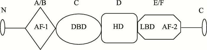
图1
PPARγ蛋白结构模式图
AF-1:激活结构域(A/B);DBD:DNA结合结构域(C);HD:铰区域(D);LBD:配体结合区域(E/F)。
Fig. 1
Schematic representation of the domain structures of PPARγ protein
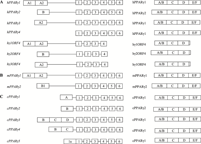
研究发现,PPARγ基因是一个多启动子调控的基因。人PPARγ基因有4个不同的启动子,由于启动子不同和选择性拼接,PPARγ基因可产生4种不同的转录异构体——hPPARγ1-4,这些转录异构体的差别仅在于5′UTR序列和长度不同[1,16](图2)。其中,转录异构体hPPARγ1、3和4都编码hPPARγ1蛋白异构体,而转录异构体hPPARγ2编码hPPARγ2蛋白异构体。最近有研究报道了人PPARγ基因的3个新转录异构体,分别命名为hγ1ORF4、hγ2ORF4和hγ3ORF4。与目前已知的人PPARγ转录本相比,这3个新转录本都缺少3′端最后2个外显子(外显子5和6)[16](图2)。其中,hγ1ORF4与hPPARγ1的5′UTR相同,hγ2ORF4和hγ3ORF4则分别与hPPARγ2和hPPARγ3的5′UTR相同(图2)。转录异构体hγ1ORF4和hγ3ORF4所编码的蛋白异构体称为hγ1ORF4,转录异构体hγ2ORF4编码的蛋白异构体称为hγ2ORF4。这两个新蛋白异构体(hγ1ORF4和hγ2ORF4)的功能与目前已知的两个蛋白异构体(hPPARγ1和hPPARγ2)不同,hPPARγ1和hPPARγ2抑制细胞增殖,而hγ1ORF4和hγ2ORF4促进细胞增殖[16,17]。小鼠PPARγ基因有2个启动子,可产生2种不同的转录异构体,分别是mPPARγ1和mPPARγ2。这两个转录异构体的5′UTR序列和长度不同,分别编码蛋白异构体mPPARγ1和mPPARγ2(图2)[14]。以前人们认为鸡PPARγ基因只有一个转录本和一个相应的蛋白异构体cPPARγ1。但最近研究发现,鸡PPARγ基因也是多启动子调控基因,它能产生5个不同的转录异构体(cPPARγ 1-5),可编码两个蛋白异构体,即cPPARγ1和cPPARγ2,其中cPPARγ2的N端比cPPARγ1的N端长6个氨基酸残基(图2)[18]。
图2
2 PPARγ基因多态性及其关联分析
PPARγ基因存在大量的单核苷酸多态性(SNP)位点,其中许多SNPs与胰岛素抵抗、肥胖症及其相关疾病相关联[19,20,21,22,23,24,25,26,27,28,29,30,31]。P12A是PPARγ基因最常见的一个SNP,该SNP与人胰岛素抵抗和2型糖尿病的发生密切相关,其中G/G(Ala/Ala)基因型可以降低人2型糖尿病的发生危险[20]。Dong等[21]在中国人群中发现,PPARγ基因C1341T位点多态性与肥胖及2型糖尿病的发生相关。另外,PPARγ基因有些SNPs还能影响PPARγ的活性和表达。人PPARγ基因P115G位点通过影响PPARγ2蛋白Ser114的磷酸化,进而改变PPARγ2的转录激活活性[22]。PPARγ基因E157D位点通过影响PPARγ与靶基因启动子区PPRE的结合,进而影响PPARγ对靶基因调控作用的改变[23]。位于PPARγ配体结合结构域的多态性位点P467L、V290M、R425C和F388L能影响PPARγ的转录激活活性[24,25]。最近,Majithia等[26]对PPARγ基因进行大规模外显子测序分析,发现PPARγ基因有49个新的SNPs,其中9个SNPs能够抑制PPARγ活性,增加糖尿病发生的危险。另外,PPARγ基因有些SNPs与动物的脂肪性状相关。李春雨等[27]在东北农业大学高、低脂双向选择系肉鸡中发现,PPARγ基因编码区有1个突变(C291T),该突变与7周龄高、低脂双向选择系肉鸡腹脂重和腹脂率显著相关。
上述SNP位点主要位于PPARγ基因外显子区域,在PPARγ基因启动子区也发现了一些SNPs。如人PPARγ2启动子区的1个SNP——rs4684847,该突变影响转录因子PRRX1与PPARγ2启动子结合,导致PPARγ2 mRNA表达下降[28]。人PPARγ3启动子C681G多态性导致STAT5B不能与PPARγ3启动子直接结合,从而抑制PPARγ基因的表达[29]。Wang等[30]发现猪PPARγ基因启动子区的2个SNPs (c.- 1633C>T和c.-1572G>A)与肌内脂肪(intramuscular fat, IMF)沉积有关。韩青等[31]在东北农业大学高、低脂双向选择系肉鸡中发现,PPARγ基因5′侧翼区 2 kb有3个SNPs,这些SNPs均与鸡腹脂重和脂腹率显著相关。
目前已在PPARγ基因的调控区和编码区发现大量SNPs,许多SNPs与人类和动物的肥胖症及其相关疾病密切相关。部分SNPs已被证明是功能性的SNPs,能够影响PPARγ基因表达和活性,导致脂肪生成、脂肪性状的改变和相关疾病的发生。但是,目前仍然有许多SNPs的作用机制尚不清楚,需要在未来进行深入研究,分析这些SNPs是如何影响PPARγ基因的表达和功能。
3 PPARγ基因遗传调控
3.1 PPARγ基因转录调控因子
脂肪细胞分化的正调控转录因子C/EBPs(CCAAT/enhancer-binding proteins)包括C/EBPα、C/EBPβ和C/EBPδ。研究表明,小鼠PPARγ1启动子区没有C/EBPs结合位点,但PPARγ2启动子区有2个C/EBPs结合位点,C/EBPα、C/EBPβ和C/EBPδ都可以结合这两个位点并促进PPARγ2表达。在脂肪细胞分化早期,C/EBPβ和C/EBPδ与PPARγ2启动子的C/EBPs位点结合,但在分化后期,C/EBPα则取代C/EBPβ和C/EBPδ而与C/EBPs位点结合,并诱导PPARγ2表达[32]。对人和小鼠的研究发现,PPARγ和C/EBPα能够相互促进彼此与染色质的结合,从而协同激活许多脂肪细胞代谢基因的表达[33]。另外,有研究发现PPARγ和TLE3也能够相互激活彼此转录,促进脂肪生成[34]。KLFs(krüppel-like factors)转录因子家族有多个成员参与脂肪生成的调控,其成员KLF4、KLF5、KLF6、KLF9和KLF15促进PPARγ的表达[35]。在小鼠3T3-L1细胞脂肪生成过程中,KLF4、KLF5和KLF6在分化早期表达,KLF9和KLF15则分别在分化中期和分化后期表达。KLF9和KLF15激活PPARγ2启动子,促进其表达[35]。目前已证明KLF5、KLF9和KLF15直接结合于PPARγ2启动子,激活PPARγ的表达[7]。
EBF1(early B-cell factor 1)也是脂肪细胞分化的一个正调控因子,直接结合于小鼠PPARγ1启动子上,促进PPARγ基因的表达[36]。转录因子SREBP1 (sterol regulatory element-binding protein-1)和E2F1 (E2F transcription factor 1)也能直接结合小鼠PPARγ1启动子,促进PPARγ基因表达[37,38]。在脂肪生成过程中,核因子NFIA(nuclear factor 1 A)表达量显著上升,敲除NFIA能抑制3T3-L1细胞的分化。研究证实,在小鼠3T3-L1细胞中NFIA直接结合于PPARγ2启动子,促进脂肪生成[7]。转录因子NRF2(nuclear factor E2-related factor 2)能够直接与小鼠PPARγ2启动子结合,促进PPARγ2的表达和脂肪的生成[39]。
ZFP423(zinc finger protein 423)是一个新发现的转录因子,过表达ZFP423可诱导非脂源性细胞NIHT3T3中PPARγ2表达,但不能诱导PPARγ1的表达。在小鼠3T3-L1前脂肪细胞中,ZFP423过表达促进脂肪生成,敲除ZFP423基因则会抑制脂肪生成,降低PPARγ1和PPARγ2的表达,但其调控PPARγ2的机制目前还不清楚[7]。PPARγ基因存在自调控,在小鼠3T3-L1细胞分化过程中,PPARγ1蛋白能与PPARγ2启动子上的PPRE直接结合,促进PPARγ2的表达和脂肪生成[40]。在3T3-L1细胞分化早期,STAT5A/B(Signal transducer and activator of transcription 5A/B)被激活并增强PPARγ2启动子的活性,从而促进PPARγ2基因的表达[41]。另外有研究发现Twist1(twist family bHLH transcription factor 1)、KROX-20也能促进PPARγ基因的转录表达,但它们是否直接结合PPARγ基因启动子而发挥作用目前还不清楚[42,43]。
GATA2(GATA binding protein 2)和GATA3(GATA binding protein 3)是脂肪细胞分化的负调控因子,能够抑制人和小鼠PPARγ2的表达,但是目前还不清楚GATA2/3是否是直接结合于PPARγ基因的启动子区来调控PPARγ基因的表达[7]。脂肪细胞分化负调控因子KLF2、KLF3和KLF7抑制PPARγ2表达和脂肪生成。研究证实,KLF2能直接结合小鼠PPARγ2启动子,抑制其基因表达,但是KLF3和KLF7是否直接结合于PPARγ基因的启动子区来调控PPARγ基因表达目前仍不清楚[44,45,46]。Zhang等发现鸡KLF2和KLF7能够抑制PPARγ3启动子的活性和脂肪生成,但是KLF2和KLF7是否直接调控鸡PPARγ基因的表达还不清楚[45,46,47]。E2F4能直接结合于小鼠PPARγ1启动子,抑制PPARγ1的转录和脂肪生成[37]。另外,对人和小鼠的研究发现,转录因子FOXO1(forkhead box O1)、HES1(hes family bHLH transcription factor 1)、PRRX1(paired related homeobox 1)和IRF抑制人和小鼠PPARγ基因的表达,从而抑制脂肪 生成[48,49,50,51]。
3.2 PPARγ活性调控因子
辅助共激活因子(coactivator)和辅助共抑制因子(corepressor)在基因表达调控中发挥重要作用。目前已发现它们可以通过蛋白互作调控PPARγ的活性(图3),从而影响脂肪生成。哺乳动物的研究表明,辅助共激活因子PGC-1α通过与PPARγ的DNA结合结构域和配体结合区域的蛋白互作,增强PPARγ介导的转录活性;而PGC-1β通过与PPARγ的转录激活结构域结合,提高PPARγ的转录激活活性。SRC1、SRC2及SRC3(steroidreceptor coactivator 1/2/3)通过自身N端的LXXLL结构与PPARγ的AF-2结构域相互作用,提高PPARγ配体依赖性的转录激活活性[52]。辅助共激活因子TRAP220、BAF60C (BRG1/BRM-associated factor 60c)和CCPG是PPARγ转录激活作用所必需的,在配体存在条件下,这3个辅助共激活因子与PPARγ相互作用,提高PPARγ的转录激活活性[52]。
尽管目前对PPARγ基因的转录调控研究已非常深入,但是许多问题仍有待于解决。PPARγ是多启动子调控基因,截止目前,人们只对其中部分启动子开展了转录调控分析, PPARγ基因的其他启动子的调控以及在脂肪生成中启动子的选择和启动子间的相互作用等还不清楚。PPARγ基因存在自调控, 但是其自调控的机制还不十分清楚。目前已鉴定出多个调控PPARγ活性的辅助共激活和共抑制因子,是否还存在其他调控PPARγ活性的辅助因子也还不清楚。这些问题都有待于未来做进一步的研究分析。
图3
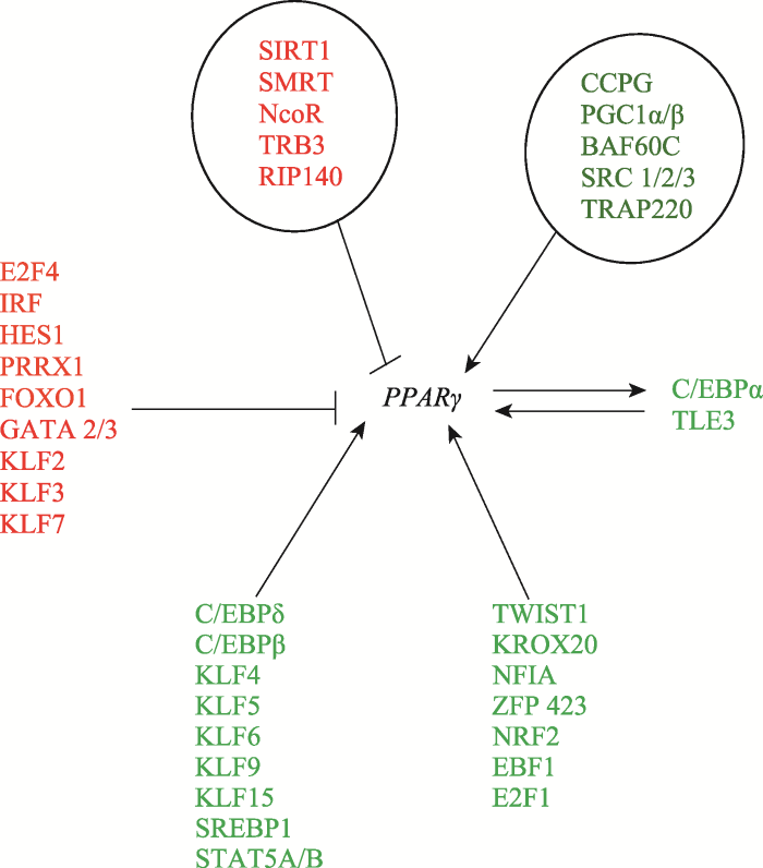
图3
PPARγ基因在脂肪生成中遗传调控
红色为抑制PPARγ基因表达的负调控转录因子,绿色为促进PPARγ基因表达的正调控转录因子。圆圈内红色字体为调控PPARγ活性的转录辅助抑制因子;圆圈内绿色字体为调控PPARγ活性的转录辅助激活因子。
Fig. 3
Genetic regulation of the PPARγ gene during adipogenesis
4 PPARγ基因表观遗传调控
4.1 DNA甲基化
Fujiki等[57]研究发现,在小鼠3T3-L1前脂肪细胞中,PPARγ2启动子高度甲基化,但随着3T3-L1前脂肪细胞的分化,PPARγ2启动子逐渐去甲基化,而PPARγ2 mRNA的表达量逐渐增加。与此相一致,应用DNA甲基化酶抑制剂(5'-aza-C)处理NIH/3T3细胞会促进PPARγ基因mRNA的表达并呈剂量依赖性。应用组蛋白去乙酰化酶抑制剂(trichostain A,TSA)处理NIH/3T3细胞,PPARγ基因mRNA的表达量没有变化。这提示,DNA甲基化在调控PPARγ基因表达中的作用高于乙酰化。比较DNA甲基化和未甲基化的PPARγ启动子的报告基因活性,发现甲基化的PPARγ启动子报告基因活性显著低于未甲基化的PPARγ启动子报告基因活性,提示DNA甲基化抑制PPARγ启动子的活性。体内研究发现,与野生小鼠相比,肥胖型糖尿病小鼠内脏脂肪细胞PPARγ2启动子的甲基化程度升高,但PPARγ2的mRNA表达下降。综上所述,无论在体内还是体外,PPARγ启动子DNA甲基化能够抑制PPARγ基因的表达,且PPARγ基因启动子DNA甲基化与肥胖症和糖尿病的发生相关。
与人和小鼠的研究类似,Sun等[58]检测了东北农业大学高、低脂双向选择系肉鸡PPARγ基因启动子的6个CpG位点(-1014、-796、-625、-548、-435和-383 bp)DNA甲基化情况及其与PPARγ表达量的相关性,结果发现,低脂鸡PPARγ3启动子的甲基化显著高于高脂鸡,而低脂鸡PPARγ基因表达量显著低于高脂鸡。这些结果提示,DNA甲基化调控鸡PPARγ基因的表达和脂肪生成。对东北农业大学高、低脂双向选择系肉鸡腹部脂肪组织DNA甲基转移酶的表达和基因组水平DNA甲基化的比较分析发现,低脂鸡腹部脂肪组织DNMT1和DNMT3A表达量均显著高于高脂鸡;与此表达结果相一致,低脂鸡腹部脂肪组织的总基因组DNA甲基化水平显著高于高脂鸡[59]。
表1 DNA甲基化调控PPARγ基因表达
Table 1
4.2 组蛋白修饰
研究发现,H3K4三甲基化(H3K4me3)能促进PPARγ基因的转录。在小鼠3T3-L1前脂肪细胞向成熟脂肪细胞分化的过程中,PPARγ2启动子区附近发生H3K4me3,该组蛋白甲基化修饰能促进PPARγ基因的表达[66]。在连续传代培养过程中,骨髓间充质干细胞的PPARγ2启动子区H3K4me3(染色质转录激活标记)与H3K27me3(染色质转录抑制标记)的比例(K4/K27)发生改变,这一变化与高代次细胞在成脂刺激剂处理时出现的PPARγ2启动子活性下降相关联[67]。在3T3-L1前脂肪和成熟脂肪细胞中,转录因子TonEB(tonicity-responsive enhancer binding protein)能够与PPARγ2启动子直接结合,使PPARγ2启动子区发生H3K9me2,从而抑制PPARγ2启动子的活性,导致PPARγ2的表达量下降,从而抑制脂肪的生成和胰岛素的敏感性[68]。
4.3 非编码RNA
非编码RNA普遍存在于真核生物中,是一类重要的转录后基因表达调控因子,参与调控众多生理过程和病理过程[71]。非编码RNA包括微小RNA (miRNA)和长链非编码RNA(lncRNA)。miRNA是一类大小为20~24核苷酸的非编码RNA分子,通过与靶基因mRNA的不完全互补结合,导致靶基因mRNA降解或蛋白翻译抑制,从而调控靶基因的表达。目前已经鉴定出多个调控PPARγ基因的miRNA (表2)。人和小鼠miRNA-27a/b、miR-301a、miR-302a和miR-548d-5p能直接作用于PPARγ基因mRNA的3′UTR,抑制PPARγ蛋白的表达和脂肪细胞分化[71,72,73,74]。miRNA-130a/b能够同时直接作用于小鼠PPARγ基因的mRNA编码区和3′UTR,下调PPARγ基因的表达,负调控脂肪生成[75,76]。与之相反,miR-375、miR-103和miR-143能够增加小鼠PPARγ2基因的表达,促进脂肪分化[77,78]。
lncRNA是一类长度超过200个核苷酸的非编码RNA,能从转录和转录后水平等多层面影响基因表达。研究发现,在3T3-L1细胞分化过程中,IncRNA也能通过影响PPARγ基因的转录,从而影响脂肪生成。Chen等[79]研究发现,在3T3-L1脂肪细胞中,lncRNA U90926能够抑制PPARγ2启动子的活性,从而抑制PPARγ的表达和脂肪生成。另外,在小鼠3T3-L1细胞分化过程中,lncRNA NEAT1在PPARγ选择性剪切中发挥调控作用,干扰NEAT1的表达能够上调PPARγ2的表达[80]。Divoux等[81]发现,过表达lncRNA HOTAIR可促进PPARγ基因的表达,促进前脂肪细胞向成熟脂肪细胞的分化,但具体的作用机制还不清楚。
表2 脂肪生成中非编码RNA调控PPARγ基因表达
Table 2
| miRNA | 功能 | 实验体系 | 靶点 | 文献 |
|---|---|---|---|---|
| miRNA-27a/b | 脂肪生成 | 3T3-L1脂肪细胞,间充质干细胞,肥胖者脂肪细胞 | 3°UTR | [71] |
| miR-301a | 脂肪生成 | 3T3-L1脂肪细胞,肥胖者脂肪细胞 | 3°UTR | [72] |
| miR-302a | 脂肪生成 | 3T3-L1脂肪细胞 | 3°UTR | [73] |
| miR-548d-5p | 脂肪生成 | 间充质干细胞 | 3°UTR | [74] |
| miRNA-130a/b | 脂肪生成 | 3T3-L1脂肪细胞,肥胖者脂肪细胞 | 编码区,3°UTR | [75,76] |
| miR-375 | 脂肪生成 | 3T3-L1脂肪细胞 | [77] | |
| miR-103 | 脂肪生成 | 3T3-L1前脂肪细胞和脂肪细胞 | [78] | |
| miR-143 | 脂肪生成 | 3T3-L1前脂肪细胞和脂肪细胞 | [78] | |
| lncRNA U90926 | 脂肪生成 | 3T3-L1前脂肪细胞和脂肪细胞 | [79] | |
| lncRNA NEAT1 | 脂肪生成 | 3T3-L1前脂肪细胞和脂肪细胞 | [80] | |
| lncRNA HOTAIR | 脂肪生成 | 人腹部皮下前脂肪细胞 | [81] |
注:箭头向下表示抑制脂肪生成,箭头向上表示促进脂肪生成。
4.4 染色质重塑
染色质重塑既可以激活基因表达,也可抑制基因表达。在3T3-L1前脂肪细胞诱导分化后的几个小时内,PPARγ基因座发生染色质重塑,染色质呈开放状态[82],其中PPARγ2启动子区是染色质开放区域之一。PPARγ2启动子区的重塑和开放是脂肪组织的特性,且依赖于PKA(protein kinase A)。研究表明,利用shRNA敲除PKA会导致PPARγ2启动子区染色质的可接近性(chromatin accessibility)下降。研究表明,SWI/SNF染色质重塑复合物调控脂肪生成中PPARγ2的表达。Brg1是SWI/SNF染色质重塑复合物的一个成分,Brg1的显性突变体抑制PPARγ、C/EBPα、C/EBPβ所诱导的成纤维细胞向脂肪细胞的转分化[7,83]。在3T3-L1前脂肪细胞分化早期,C/EBPs蛋白与PPARγ2启动子结合,随后聚合酶Ⅱ及基础转录因子组装于PPARγ2启动子区,最后SWI/ SNF染色质重塑复合物和转录因子TFIIH才组装于PPARγ2启动子上,促进转录起始复合物的形成[7]。
综上所述,表观遗传在调控PPARγ基因表达和脂肪生成中发挥重要的作用。但PPARγ基因的表观遗传调控研究刚刚起步,许多表观遗传调控的作用机制尚不清楚。例如,PPARγ基因各启动子的表观遗传调控机制是否存在差异,DNA甲基化、组蛋白修饰、染色质重塑等表观遗传调控方式如何协同调控PPARγ基因的表达。此外,许多PPARγ基因的转录因子如E2F1、NRF1、Myc、SP1等,它们的DNA结合区都有CpG位点,但是PPARγ基因启动子区的DNA甲基化是否影响这些转录因子与启动子的结合,这些问题都还有待于进一步深入探索和研究。
5 展望
目前人们对脂肪生成中PPARγ基因调控的认识还不够,还有许多问题有待于研究。由于PPARγ在人和动物脂肪生成和脂肪发育以及动物生产中的重要性,PPARγ基因的研究仍将是脂肪生物学、医学和动物遗传学的研究热点。近年来,高通量分析和质谱分析等技术发展迅速,已成为研究基因遗传和表观遗传调控的重要工具。相信借助于这些新技术以及研究的不断深入,PPARγ基因在脂肪生成中的遗传和表观遗传调控机制将被逐步阐明,这将有助于人类最终了解脂肪生成和脂肪生长发育的遗传和表观遗传机制,也将有助于推动以PPARγ基因为靶点的肥胖症及其相关疾病的药物开发。另外,也可尝试采用PPARγ基因遗传标记(如功能性SNP)和表观遗传标记(如DNA甲基化标记)开展畜禽的分子标记辅助育种,加速优质低脂畜禽新品种的培育。
(责任编委: 蒋思文)
参考文献
PPARγ and its role in cardiovascular diseases
Three important transcription factors related to lipogenesis and adipogenesis in mam mal
Characterization of chicken PPARγ expression and its impact on adipocyte proliferation and differentiation
为分析鸡PPARγ基因的组织表达特性及其在脂肪细胞增殖和分化过程中的功能, 文章以东北农业大学高、低腹脂双向选择品系肉鸡为实验材料, 利用Western blotting方法, 检测PPARγ基因的组织表达特性及其在高、低脂系肉鸡腹部脂肪组织间的表达差异; 采用RNAi技术, 在鸡原代脂肪细胞中抑制PPARγ基因的表达后, 通过MTT和油红O提取比色的方法, 研究鸡PPARγ基因对脂肪细胞增殖和分化的调控作用; 利用Real-time PCR和Western blotting技术, 分析PPARγ基因表达下调后, 其他脂肪细胞分化转录因子以及与脂肪细胞分化相关的重要基因的表达变化情况。结果表明, PPARγ基因在7周龄高脂系肉鸡腹部脂肪组织、肌胃、脾脏、肾脏组织中表达量较高, 在心脏中表达量较低, 在肝脏、胸肌、腿肌、十二指肠中未检测到表达信号; 与高脂系相比, PPARγ基因在5和7周龄低脂系肉鸡腹部脂肪组织中的表达量较低(P<0.05); PPARγ基因的表达量下降后, 鸡脂肪细胞的增殖能力增强, 分化能力减弱; 同时, C/EBPα、SREBP1、A-FABP、Perilipin1、LPL、IGFBP-2基因的表达量均下降(P<0.05)。由此可见, PPARγ基因的表达可能与肉鸡腹部脂肪的沉积有一定的关系, 该基因可能是调控鸡脂肪细胞增殖与分化的关键因子。
鸡PPARγ基因的表达特性及其对脂肪细胞增殖分化的影响
为分析鸡PPARγ基因的组织表达特性及其在脂肪细胞增殖和分化过程中的功能, 文章以东北农业大学高、低腹脂双向选择品系肉鸡为实验材料, 利用Western blotting方法, 检测PPARγ基因的组织表达特性及其在高、低脂系肉鸡腹部脂肪组织间的表达差异; 采用RNAi技术, 在鸡原代脂肪细胞中抑制PPARγ基因的表达后, 通过MTT和油红O提取比色的方法, 研究鸡PPARγ基因对脂肪细胞增殖和分化的调控作用; 利用Real-time PCR和Western blotting技术, 分析PPARγ基因表达下调后, 其他脂肪细胞分化转录因子以及与脂肪细胞分化相关的重要基因的表达变化情况。结果表明, PPARγ基因在7周龄高脂系肉鸡腹部脂肪组织、肌胃、脾脏、肾脏组织中表达量较高, 在心脏中表达量较低, 在肝脏、胸肌、腿肌、十二指肠中未检测到表达信号; 与高脂系相比, PPARγ基因在5和7周龄低脂系肉鸡腹部脂肪组织中的表达量较低(P<0.05); PPARγ基因的表达量下降后, 鸡脂肪细胞的增殖能力增强, 分化能力减弱; 同时, C/EBPα、SREBP1、A-FABP、Perilipin1、LPL、IGFBP-2基因的表达量均下降(P<0.05)。由此可见, PPARγ基因的表达可能与肉鸡腹部脂肪的沉积有一定的关系, 该基因可能是调控鸡脂肪细胞增殖与分化的关键因子。
Functional analysis of the chicken PPARγ gene 5'-flanking region and C/EBPα- mediated gene regulation
Peroxisome proliferator-activated receptor-γ ( PPARγ) and CCAAT/enhancer binding protein-α (C/EBPα) are the master regulators of adipogenesis. The regulatory mechanism of PPARγ and C/EBPα gene expression is clear in mammals, however, little is known in chicken. The aim of the present study was to characterize chicken PPARγ promoter and investigate whether PPARγ could be regulated by C/EBPα in chickens. A 2-kb nucleotide sequence upstream of the start codon of chicken PPARγ gene was cloned and characterized by using bioinformatics and experimental approaches. This 2-kb promoter region exhibited strong promoter activity in DF1 cells. The reporter gene assay showed that the chicken C/EBPα could activate PPARγ gene promoter. Further study by electrophoretic mobility shift assay and mutational analysis revealed that the chicken C/EBPα could directly bind to and regulate the PPARγ gene promoter. Our results demonstrate that PPARγ can be directly regulated by C/EBPα in chickens .
Molecular regulation of adipogenesis
PPARγ: a nuclear regulator of metabolism, differentiation, and cell growth
The perexiseme proliferator-activated receptors (PPARs) comprise an important subfamily of the nuclear hormone receptor (NHR) superfamily. These ligand-activated transcription factors have been intensively studied for more than a decade and have been implicated in such diverse pathways as lipid and glucose homeostasis, control of cellular proliferation, and differentiation. The name PPAR derives from the initial cloning of one isoform as a target of various xenobiotic compounds that were observed to induce proliferation of peroxisomes in the liver (1). This protein was called the peroxisome proliferator-activated receptor, now known as PPAR#alpha#. Within a few years, the group of PPARs was expanded to include PPAR#gamma# and PPAR#delta# (also referred to as PPAR#beta#, NUC1, and FAa, R) (2-6). This review will focus on PPAR#gamma#.
Transcriptional and epigenetic regulation of PPARγ expression during adipogenesis
URL
PMID:4046494
[本文引用: 10]

The nuclear receptor PPAR?? is a master regulator of adipogenesis. PPAR?? is highly expressed in adipose tissues and its expression is markedly induced during adipogenesis. In this review, we describe the current knowledge, as well as future directions, on transcriptional and epigenetic regulation of PPAR?? expression during adipogenesis. Investigating the molecular mechanisms that control PPAR?? expression during adipogenesis is critical for understanding the development of white and brown adipose tissues, as well as pathological conditions such as obesity and diabetes. The robust induction of PPAR?? expression during adipogenesis also serves as an excellent model system for studying transcriptional and epigenetic regulation of cell-type-specific gene expression.
PPARγ is required for placental, cardiac, and adipose tissue development
Abstract The nuclear hormone receptor PPAR gamma promotes adipogenesis and macrophage differentiation and is a primary pharmacological target in the treatment of type II diabetes. Here, we show that PPAR gamma gene knockout results in two independent lethal phases. Initially, PPAR gamma deficiency interferes with terminal differentiation of the trophoblast and placental vascularization, leading to severe myocardial thinning and death by E10.0. Supplementing PPAR gamma null embryos with wild-type placentas via aggregation with tetraploid embryos corrects the cardiac defect, implicating a previously unrecognized dependence of the developing heart on a functional placenta. A tetraploid-rescued mutant surviving to term exhibited another lethal combination of pathologies, including lipodystrophy and multiple hemorrhages. These findings both confirm and expand the current known spectrum of physiological functions regulated by PPAR gamma.
PPAR gamma in neuroblastoma: the translational perspectives of hypoglycemic drugs
Neuroblastoma (NB) is the most common and aggressive pediatric cancer, characterized by a remarkable phenotypic diversity and high malignancy. The heterogeneous clinical behavior, ranging from spontaneous remission to fatal metastatic disease, is attributable to NB biology and genetics. Despite major advances in therapies, NB is still associated with a high morbidity and mortality. Thus, novel diagnostic, prognostic, and therapeutic approaches are required, mainly to improve treatment outcomes of high-risk NB patients. Among neuroepithelial cancers, NB is the most studied tumor as far as PPAR ligands are concerned. PPAR ligands are endowed with antitumoral effects, mainly acting on cancer stem cells, and constitute a possible add-on therapy to antiblastic drugs, in particular for NB with unfavourable prognosis. While discussing clinical background, this review will provide a synopsis of the major studies about PPAR expression in NB, focusing on the potential beneficial effects of hypoglycemic drugs, thiazolidinediones and metformin, to reduce the occurrence of relapses as well as tumor regrowth in NB patients.
Revisiting PPARγ as a target for the treatment of metabolic disorders
As the prevalence of obesity has increased explosively over the last several decades, associated metabolic disorders, including type 2 diabetes, dyslipidemia, hypertension, and cardiovascular diseases, have been also increased. Thus, new strategies for preventing and treating them are needed. The nuclear peroxisome proliferator-activated receptors (PPARs) are involved fundamentally in regulating energy homeostasis; thus, they have been considered attractive drug targets for addressing metabolic disorders. Among the PPARs, PPAR纬 is a master regulator of gene expression for metabolism, inflammation, and other pathways in many cell types, especially adipocytes. It is a physiological receptor of the potent anti-diabetic drugs of the thiazolidinediones (TZDs) class, including rosiglitazone (Avandia). However, TZDs have undesirable and severe side effects, such as weight gain, fluid retention, and cardiovascular dysfunction. Recently, many reports have suggested that PPAR纬 could be modulated by post-translational modifications (PTMs), and modulation of PTM has been considered as novel approaches for treating metabolic disorders with fewer side effects than the TZDs. In this review, we discuss how PTM of PPAR纬 may be regulated and issues to be considered in making novel anti-diabetic drugs that can modulate the PTM of PPAR纬. [BMB Reports 2014; 47(11): 599-608]
Peroxisome proliferator-activated receptor-γ is critical to cardiac Fibrosis
Peroxisome proliferator-activated receptor-?? (PPAR??) is a ligand-activated transcription factor belonging to the nuclear receptor superfamily, which plays a central role in regulating lipid and glucose metabolism. However, accumulating evidence demonstrates that PPAR?? agonists have potential to reduce inflammation, influence the balance of immune cells, suppress oxidative stress, and improve endothelial function, which are all involved in the cellular and molecular mechanisms of cardiac fibrosis. Thus, in this review we discuss the role of PPAR?? in various cardiovascular conditions associated with cardiac fibrosis, including diabetes mellitus, hypertension, myocardial infarction, heart failure, ischemia/reperfusion injury, atrial fibrillation, and several other cardiovascular disease (CVD) conditions, and summarize the developmental status of PPAR?? agonists for the clinical management of CVD.
Peroxisome proliferator-activated receptors and the heart: lessons from the past and future directions
Peroxisome proliferator-activated receptors (PPARs) belong to the nuclear family of ligand activated transcriptional factors and comprise three different isoforms, PPAR-??, PPAR-??/??, and PPAR-??. The main role of PPARs is to regulate the expression of genes involved in lipid and glucose metabolism. Several studies have demonstrated that PPAR agonists improve dyslipidemia and glucose control in animals, supporting their potential as a promising therapeutic option to treat diabetes and dyslipidemia. However, substantial differences exist in the therapeutic or adverse effects of specific drug candidates, and clinical studies have yielded inconsistent data on their cardioprotective effects. This review summarizes the current knowledge regarding the molecular function of PPARs and the mechanisms of the PPAR regulation by posttranslational modification in the heart. We also describe the results and lessons learned from important clinical trials on PPAR agonists and discuss the potential future directions for this class of drugs.
The organization, promoter analysis, and expression of the human PPARγ gene
Structural organization of mouse peroxisome proliferator-activated receptor gamma (mPPAR gamma) gene: alternative promoter use and different splicing yield two mPPAR gamma isoforms
To gain insight into the regulation of expression of peroxisome proliferator-activated receptor (PPAR) isoforms, we have determined the structural organization of the mouse PPAR γ (mPPARγ) gene. This gene extends >105 kb and gives rise to two mRNAs (mPPARγ1 and mPPARγ2) that differ at their 5' ends. The mPPARγ2 cDNA encodes an additional 30 amino acids N-terminal to the first ATG codon of mPPARγ1 and reveals a different 5' untranslated sequence. We show that mPPARγ1 mRNA is encoded by eight exons, whereas the mPPARγ2 mRNA is encoded by seven exons. Most of the 5' untranslated sequence of mPPARγ1 mRNA is encoded by two exons, whereas the 5' untranslated sequence and the extra 30 N-terminal amino acids of mPPARγ2 are encoded by one exon, which is located between the second and third exons coding for mPPARγ1. The last six exons of mPPARγ gene code for identical sequences in mPPARγ1 and mPPARγ2 isoforms. The mPPARγ1 and mPPARγ2 isoforms are transcribed from different promoters. The mPPARγ gene has been mapped to chromosome 6 E3-F1 by in situ hybridization using a biotin-labeled probe. These results establish that at least one of the PPAR genes yields more than one protein product, similar to that encountered with retinoid X receptor and retinoic acid receptor genes. The existence of multiple PPAR isoforms transcribed from different promoters could increase the diversity of ligand and tissue-specific transcriptional responses.
A novel splicing variant of peroxisome proliferator-activated receptor-γ ( Pparγ1sv) cooperatively regulates adipocyte differentiation with Pparγ2
D'Esposito V, Beguinot F, Formisano P, Costa V, Ciccodicola A PPARG in human adipogenesis: differential contribution of canonical transcripts and dominant negative isoforms
Peroxisome proliferator activated receptors at the crossroad of obesity, diabetes, and pancreatic cancer
Pancreatic ductal adenocarcinoma(PDAC) is the fourth cause of cancer death with an overall survival of 5% at five years. The development of PDAC is characteristically associated to the accumulation of distinctive genetic mutations and is preceded by the exposure to several risk factors. Epidemiology has demonstrated that PDAC risk factors may be non-modifiable risks(sex, age, presence of genetic mutations, ethnicity) and modifiable and co-morbidity factors related to the specific habits and lifestyle. Recently it has become evident that obesity and diabetes are two important modifiable risk factors for PDAC. Obesity and diabetes are complex systemic and intertwined diseases and, over the years, experimental evidence indicate that insulin-resistance, alteration of adipokines, especially leptin and adiponectin, oxidative stress and inflammation may play a role in PDAC. Peroxisome proliferator activated receptor-纬(PPAR纬) is a nuclear receptor transcription factor that is implicated in the regulation of metabolism, differentiation and inflammation. PPAR纬 is a key regulator of adipocytes differentiation, regulates insulin and adipokines production and secretion, may modulate inflammation, and it is implicated in PDAC. PPAR纬 agonists are used in the treatment of diabetes and oxidative stressassociated diseases and have been evaluated for the treatment of PDAC. PPAR纬 is at the cross-road of diabetes, obesity, and PDAC and it is an interesting target to pharmacologically prevent PDAC in obese and diabetic patients.
Identification and characterization of transcript variants of chicken peroxisome proliferator-activated receptor gamma
Peroxisome proliferator-activated receptor gamma regulates adipogenesis. The genomic structure of the chicken peroxisome proliferator-activated receptor gamma (cPPARγ) gene has not been fully characterized, and only one cPPARγ gene mRNA sequence has been reported in genetic databases. Using 5' rapid amplification of cDNA ends, we identified five different cPPARγ mRNAs that are transcribed from three transcription initiation sites. The open reading frame analysis showed that these five cPPARγ transcript variants (cPPARγ1 to 5) could encode two cPPARγ protein isoforms (cPPARγ1 and cPPARγ2), which differ only in their N-terminal region. Quantitative real-time RT-PCR analysis showed that, of these five cPPARγ transcript variants, cPPARγ1 was ubiquitously highly expressed in various chicken tissues, including adipose tissue, liver, kidney, spleen and duodenal; cPPARγ2 was exclusively highly expressed in adipose tissue; cPPARγ3 was highly expressed in adipose tissue, kidney, spleen and liver; cPPARγ4 and cPPARγ5 were ubiquitously weakly expressed in all the tested tissues, and comparatively, cPPARγ5 was highly expressed in adipose tissue, heart, liver and kidney. The comparison of the expression of the five cPPARγ transcript variants showed that adipose tissue cPPARγ1 expression was significantly higher in the fat line than in the lean line from 2 to 7 wk of age (P<0.05 or P<0.01). Adipose tissue cPPARγ3 expression was significantly higher in the fat line than in the lean line at 3, 5 and 6 wk of age (P<0.01, P<0.05), but lower at 4 wk of age (P<0.05). Adipose tissue cPPARγ5 expression was significantly higher in the fat line than in the lean line at 3, 4, and 6 wk of age (P<0.01) and at 2 and 7 wk of age (P<0.05). This is the first report of transcript variants and protein isoforms of cPPARγ gene. Our findings provided a foundation for future investigations of the function and regulation of cPPARγ gene in adipose tissue development.
Familial partial lipodystrophy phenotype resulting from a single-base mutation I n deoxyribonucleic acid-binding domain of peroxisome proliferator-activated receptor-γ
Familial partial lipodystrophy (FPLD) results from coding sequence mutations either in LMNA, encoding nuclear lamin A/C, or in PPARG, encoding peroxisome proliferator-activated receptor-gamma (PPARgamma). The LMNA form is called FPLD2 (MIM 151660) and the PPARG form is called FPLD3 (MIM 604367).Our objective was to investigate whether the clinical phenotype of this proband is due to mutation(s) in PPARgamma.This is a case report. Patient and Setting: A 31-yr-old female with the clinical phenotype of FPLD3, i.e. lipodystrophy and early childhood diabetes with extreme insulin resistance and hypertriglyceridemia leading to recurrent pancreatitis, was assessed at an academic medical center.The proband was heterozygous for a novel C-->T mutation in the PPARG gene that led to the substitution of arginine 194 in PPARgamma2 isoform, a conserved residue located in the zinc finger structure involved in DNA binding, by tryptophan (R194W). The mutation was absent from the genomes of 100 healthy Caucasians. In vitro analysis of the mutated protein showed that R194W (and R166W in PPARgamma1 isoform) could not bind to DNA and had no transcriptional activity. Furthermore, R194W had no dominant-negative activity.The R194W mutation in PPARG disrupts its DNA binding activity and through haploinsufficiency leads to clinical manifestation of FPLD3 and the associated metabolic disturbances.
Association of polymorphisms of peroxisome proliferator activated receptors in early and late onset of type 2 diabetes mellitus
There is a strong association of PPAR γ, PPAR α and PPAR δ genes on the susceptibility of T2DM in late onset but not with the early onset of T2DM subjects in North Indian Population: Dual association of PPAR γ was observed with its genotype G/G (Ala/Ala) favoring protection against T2DM and genotype C/C (Pro/Pro) favoring susceptibility to T2DM. Association of intron7 polymorphism of PPAR α and +T294C polymorphism of PPAR δ on the susceptibility to T2DM requires further analysis.
Role of peroxisome proliferator-activated receptors gene polymorphisms in type 2 diabetes and metabolic syndrome
Metabolic syndrome(MetS) and type 2 diabetes mellitus(T2DM) are the serious public health problems worldwide.Moreover,it is estimated that MetS patients have about five-fold greater risk of the T2 DM development compared with people without the syndrome.Peroxisome proliferator-activated receptors are a subgroup of the nuclear hormone receptor superfamily of ligand-activated transcription factors which play an important role in the pathogenesis of MetS and T2 DM.All three members of the peroxisome proliferator-activated receptor(PPAR) nuclear receptor subfamily,PPARα,PPARp/5 and PPARγ are critical in regulating insulin sensitivity,adipogenesis,lipid metabolism,and blood pressure.Recently,more and more studies indicated that the gene polymorphism of PPARs,such as Leu~(162)Val and Val~(227)Ala of PPARα,+294T C of PPARβ/δ,Pro~(12)Ala and C1431 T of PPARγ,are significantly associated with the onset and progressing of MetS and T2 DM in different population worldwide.Furthermore,a large body of evidence demonstrated that the glucose metabolism and lipid metabolism were influenced by gene-gene interaction among PPARs genes.However,given the complexity pathogenesis of metabolic disease,it is unlikely that genetic variation of a single locus would provide an adequate explanation of inter-individual differences which results in diverse clinical syndromes.Thus,gene-gene interactions and gene-environment interactions associated with T2 DM and MetS need future comprehensive studies.
Obesity associated with a mutation in a genetic regulator of adipocyte differentiation
PPARγ mutations, lipodystrophy and diabetes
The focus of this review is the lipodystrophy syndrome caused by mutation in the PPARγ nuclear receptor – partial familial lipodystrophy FPLD3. To provide a broader context for how these mutations act to generate the clinical features of partial lipodystrophy we will review the basic biology of PPARγ and also survey the set PPARγ genetic variants that do not cause lipodystrophy, but are nonetheless associated with clinically related syndromes, specifically type 2 diabetes.
O'Rahilly S, Chatterjee K. Non-DNA binding, dominant-negative, human PPARγ mutations cause lipodystrophic insulin resistance
O'Rahilly S. Dominant negative mutations in human PPARγ associated with severe insulin resistance, diabetes mellitus and hypertension
GoT2D consortium, NHGRI JHS/FHS allelic spectrum project, SIGMA T2D consortium, T2D-GENES consortium, Rosen ED, Altshuler D. Rare variants in PPARG with decreased activity in adipocyte differentiation are associated with increased risk of type 2 diabetes
The study of SNPs in chicken PPARγ gene and their associations with fatness trait. Progress in animal genetic breeding in China Progress——The thirteenth national symposium on animal genetics breeding in China
鸡PPARγ基因SNPs与脂肪性状相关的研究. 见:中国动物遗传育种研究进展——第十三次全国动物遗传育种学术讨论会论文集
Leveraging cross-species transcription factor binding site patterns: from diabetes risk loci to disease mechanisms
Genome-wide association studies have revealed numerous risk loci associated with diverse diseases. However, identification of disease-causing variants within association loci remains a major challenge. Divergence in gene expression due to cis -regulatory variants in noncoding regions is central to disease susceptibility. We show that integrative computational analysis of phylogenetic conservation with a complexity assessment of co-occurring transcription factor binding sites (TFBS) can identify cis -regulatory variants and elucidate their mechanistic role in disease. Analysis of established type 2 diabetes risk loci revealed a striking clustering of distinct homeobox TFBS. We identified the PRRX1 homeobox factor as a repressor of PPARG2 expression in adipose cells and demonstrate its adverse effect on lipid metabolism and systemic insulin sensitivity, dependent on the rs4684847 risk allele that triggers PRRX1 binding. Thus, cross-species conservation analysis at the level of co-occurring TFBS provides a valuable contribution to the translation of genetic association signals to disease-related molecular mechanisms.
A functional polymorphism in a STAT5B site of the human PPARγ3 gene promoter affects height and lipid metabolism in a French population
ABSTRACT The peroxisome proliferator-activated receptor-gamma (PPARgamma) plays a role in adipocyte differentiation and insulin sensitization. It has been shown that genetic variation in the PPARgamma gene alters body weight control, lipid and insulin homeostasis, and the susceptibility to type 2 diabetes. Four PPARgamma isoforms are generated by alternative splicing and promoter usage. PPARgamma3 is only expressed in adipose tissue, colon, and macrophages and therefore seems to be a good candidate gene for metabolic and cardiovascular-associated diseases. In the present study, we looked for genetic variation in the PPARgamma3 promoter. The proximal PPARgamma3 promoter was sequenced in 20 individuals. We detected a C/G polymorphism at position -681 from exon A2. Interestingly, it was located in a signal transducer and activator of transcription 5B (STAT5B) binding consensus site. In a French population (n=836), the -681G allele was associated with increased height and plasma low-density lipoprotein cholesterol concentrations. In vitro, we showed that the -681G allele completely abolished the binding of STAT5B to the cognate promoter element as well as the transactivation of the PPARgamma3 promoter by the growth hormone/STAT5B pathway. Our results suggest that PPARgamma3 may regulate the control of height and lipid homeostasis via the STAT5B pathway.
Two completely linked polymorphisms in the PPARG transcriptional regulatory region significantly affect gene expression and intramuscular fat deposition in the longissimus dorsimuscle of Erhualian pigs
Correlation between peroxisome proliferator-activated receptor g 5'-flanking region haplotypes with the growth and body composition traits in chickens
PPARγ基因5′侧翼区单倍型与鸡生长和体组成性状的相关研究
C/EBPα induces adipogenesis through PPARγ: a unified pathway
PPARγ and C/EBPα are critical transcription factors in adipogenesis, but the precise role of these proteins has been difficult to ascertain because they positively regulate each other's expression. Questions remain about whether these factors operate independently in separate, parallel pathways of differentiation, or whether a single pathway exists. PPARγ can promote adipogenesis in C/EBPα-deficient cells, but the converse has not been tested. We have created an immortalized line of fibroblasts lacking PPARγ, which we use to show that C/EBPα has no ability to promote adipogenesis in the absence of PPARγ. These results indicate that C/EBPα and PPARγ participate in a single pathway of fat cell development with PPARγ being the proximal effector of adipogenesis.
Peroxisome proliferator-activated receptor γ and C/EBPα synergistically activate key metabolic adipocyte genes by assisted loading
Peroxisome proliferator-activated receptor γ (PPARγ) and CCAAT/enhancer binding protein α (C/EBPα) are key activators of adipogenesis. They mutually induce the expression of each other and have been reported to cooperate in activation of a few adipocyte genes. Recently, genome-wide profiling revealed a high degree of overlap between PPARγ and C/EBPα binding in adipocytes, suggesting that cooperativeness could be mediated through common binding sites. To directly investigate the interplay between PPARγ and C/EBPα at shared binding sites, we established a fibroblastic model system in which PPARγ and C/EBPα can be independently expressed. Using RNA sequencing, we demonstrate that coexpression of PPARγ and C/EBPα leads to synergistic activation of many key metabolic adipocyte genes. This is associated with extensive C/EBPα-mediated reprogramming of PPARγ binding and vice versa in the vicinity of these genes, as determined by chromatin immunoprecipitation combined with deep sequencing. Our results indicate that this is at least partly mediated by assisted loading involving chromatin remodeling directed by the leading factor. In conclusion, we report a novel mechanism by which the key adipogenic transcription factors, PPARγ and C/EBPα, cooperate in activation of the adipocyte gene program.
TLE3 is a dual-function transcriptional coregulator of adipogenesis
78 High-throughput cDNA screening identified TLE3 as a promoter of adipocyte differentiation 78 TLE3 is induced by PPARγ and participates in a feed-forward adipogenic loop 78 TLE3 facilitates PPARγ action on the promoters of adipocyte target genes 78 TLE3 blocks β-catenin action and antagonizes Wnt signaling during adipogenesis
Study on adipocyte differentiation molecular mechanisms
白色脂肪细胞分化分子机制研究进展
Critical role for Ebf1 and Ebf2 in the adipogenic transcriptional cascade
The Ebf (O/E) family of helix-loop-helix transcription factors plays a significant role in B lymphocyte and neuronal development. The three primary members of this family, Ebf1, 2, and 3, are all expressed in adipocytes, and Ebf1 promotes adipogenesis when overexpressed in NIH 3T3 fibroblasts. Here we report that these three proteins have adipogenic potential in multiple cellular models and that peroxisome proliferator-activated receptor gamma (PPAR gamma) is required for this effect, at least in part due to direct activation of the PPAR gamma 1 promoter by Ebf1. Ebf1 also directly binds to and activates the C/EBP alpha promoter, which exerts positive feedback on C/EBP delta expression. Despite this, C/EBP alpha is dispensable for the adipogenic action of Ebf proteins. Ebf1 itself is induced by C/EBP beta and delta, which bind and activate its promoter. Reduction of Ebf1 and Ebf2 proteins by specific short hairpin RNA blocks differentiation of 3T3-L1 cells, suggesting a critical role for these factors and the absence of functional redundancy between members of this family. Altogether, these data place Ebf1 within the known transcriptional cascade of adipogenesis and suggest critical roles for Ebf1 and Ebf2.
E2Fs regulate adipocyte differentiation
When preadipocytes reenter the cell cycle, PPARγ expression is induced, coincident with an increase in DNA synthesis, suggesting the involvement of the E2F family of cell cycle regulators. We show here that E2F1 induces PPARγ transcription during clonal expansion, whereas E2F4 represses PPARγ expression during terminal adipocyte differentiation. Using a combination of in vivo experiments with knockout and chimeric animals and in vitro experiments, we demonstrate that the absence of E2F1 impairs, whereas depletion of E2F4 stimulates, adipogenesis. E2Fs hence represent the link between proliferative signaling pathways, triggering clonal expansion, and terminal adipocyte differentiation through regulation of PPARγ expression. This underscores the complex role of the E2F protein family in the control of both cell proliferation and differentiation.
Regulation of peroxisome proliferator-activated receptor γ expression by adipocyte differentiation and determination factor 1/sterol regulatory element binding protein 1: implications for adipocyte differentiation and metabolism
Suppression of Nrf2 attenuates adipogenesis and decreases FGF21 expression through PPAR gamma in 3T3-L1 cells
Adipogenesis is the process of differentiation from preadipocytes to adipocytes and is orchestrated by various transcription factors, such as the peroxisome proliferator-activated receptor gamma (PPARγ) and the CCAAT-enhancer-binding protein alpha (C/EBPα). Oxidative stress is also a crucial factor in adipogenesis, and adipocyte differentiation is affected by the cellular redox status. The nuclear factor E2-related factor 2 (Nrf2), which is a basic leucine zipper (bZIP) transcription factor, acts as a regulator of cellular oxidative stress. Although several previous studies examined the function of Nrf2 in adipogenesis, their results were controversial. In this study, we investigated whether the suppression of Nrf2 in 3T3-L1 cells affected adipogenesis. We found that adipogenesis master regulator genes, such as PPARγ and C/EBPα , were downregulated during the differentiation stage in Nrf2-knockdown 3T3-L1 cells. Moreover, the fibroblast growth factor 21 (FGF21) and manganese superoxide dismutase (MnSOD) were markedly downregulated in Nrf2-knockdown 3T3-L1 cells. Taken together, the results of the present study suggest that the suppression of Nrf2 attenuates adipogenesis and decreases FGF21 expression through PPARγ in 3T3-L1 cells.
Activation of PPARγ2 by PPARγ1 through a functional PPRE in transdifferentiation of myoblasts to adipocytes induced by EPA
PPARγ and Wnt signaling are central positive and negative regulators of adipogenesis, respectively. Here we identified that, eicosapentaenoic acid (EPA) could effectively induce the transdifferentiation of myoblasts into adipocytes through modulation of both PPARγ expression and Wnt signaling. During the early stage of transdifferentiation, EPA activates PPARδ and PPARγ1, which in turn targets β-catenin to degradation and down-regulates Wnt/β-catenin signaling, such that the myogenic fate of myoblasts could be switched to adipogenesis. In addition, EPA up-regulates the expression of PPARγ1 by activating RXRα, then PPARγ1 binds to the functional peroxisome proliferator responsive element (PPRE) in the promoter of adipocyte-specific PPARγ2 to continuously activate the expression of PPARγ2 throughout the transdifferentiation process. Our data indicated that EPA acts as a dual-function stimulator of adipogenesis that both inhibits Wnt signaling and induces PPARγ2 expression to facilitate the transdifferentiation program, and the transcriptional activation of PPARγ2 by PPARγ1 is not only the key factor for the transdifferentiation of myoblasts to adipocytes, but also the crucial evidence for successful transdifferentiation. The present findings provided insight for the first time as to how EPA induces the transdifferentiation of myoblasts to adipocytes, but also provide new clues for strategies to prevent and treat some metabolic diseases.
Stimulation of 3T3- L1 adipogenesis by signal transducer and activator of transcription 5
Signal transducer and activator of transcription 5 () mediates of many cytokines and . Here we show that functions as an initial mediator of adipogenesis. The preadipocyte line 3T3-L1 undergoes upon appropriate hormonal induction. We found that Stat5A and Stat5B were strongly activated at an early stage of 3T3-L1 differentiation. To investigate physiological roles of in adipogenesis, we have constructed 3T3-L1 lines in which either an exogenous wild type (wt) or dominant negative (dn) form of Stat5A expression was controlled under the -regulatable promoter. Precocious induction of wt-Stat5A in promoted accumulation of within the . In contrast, induction of dn-Stat5A attenuated accumulation. Northern blot analyses revealed that the expression of proadipogenic transcription factors was influenced in a complementary fashion by ectopic expression of either wt- or dn-Stat5A. Notably, regulated expression of , which plays crucial roles in adipogenesis. We have also generated in which dn-Stat5A is expressed in an adipose tissue-specific fashion and found attenuation of and of many adipocyte-related genes. These results highlight a novel role of in .
The role and regulation of transcription factors Twist 1 and PPARγ in mice 3T3-L1 adipocytes
转录因子Twist 1和PPARγ在3T3-L1细胞中的作用及调控关系
Transcriptional control of adipocyte formation
A detailed understanding of the processes governing adipose tissue formation will be instrumental in combating the obesity epidemic. Much progress has been made in the last two decades in defining transcriptional events controlling the differentiation of mesenchymal stem cells into adipocytes. A complex network of transcription factors and cell-cycle regulators, in concert with specific transcriptional coactivators and corepressors, respond to extracellular stimuli to activate or repress adipocyte differentiation. This review summarizes advances in this field, which constitute a framework for potential antiobesity strategies.
Targeted disruption of the Basic Krüppel- like Factor gene (Klf3) reveals a role in adipogenesis
Kr眉ppel-like factors (KLFs) recognize CACCC and GC-rich sequences in gene regulatory elements. Here, we describe the disruption of the murine basic Kr眉ppel-like factor gene (Bklf or Klf3). Klf3 knockout mice have less white adipose tissue, and their fat pads contain smaller and fewer cells. Adipocyte differentiation is altered in murine embryonic fibroblasts from Klf3 knockouts. Klf3 expression was studied in the 3T3-L1 cellular system. Adipocyte differentiation is accompanied by a decline in Klf3 expression, and forced overexpression of Klf3 blocks 3T3-L1 differentiation. Klf3 represses transcription by recruiting C-terminal binding protein (CtBP) corepressors. CtBPs bind NADH and may function as metabolic sensors. A Klf3 mutant that does not bind CtBP cannot block adipogenesis. Other KLFs, Klf2, Klf5, and Klf15, also regulate adipogenesis, and functional CACCC elements occur in key adipogenic genes, including in the C/ebpalpha promoter. We find that C/ebpalpha is derepressed in Klf3 and Ctbp knockout fibroblasts and adipocytes from Klf3 knockout mice. Chromatin immunoprecipitations confirm that Klf3 binds the C/ebpalpha promoter in vivo. These results implicate Klf3 and CtBP in controlling adipogenesis.
Cloning, tissue expression and polymorphisms of chicken Krüppel- like factor 7 gene
<P>Krüppel-like factor 7 (KLF7) has been extensively studied in mammalian species, but its role in birds is still unclear. In the current study, cloning and sequencing showed that the full-length coding region of chicken KLF7 (Gallus gallus KLF7, gKLF7) was 891 bp long, encoding 296 amino acids. In addition, real-time RT-PCR analysis showed that gKLF7 was broadly expressed in all 15 chicken tissues selected, and its expression was significantly different in spleen, proventriculus, abdominal fat, brain, leg muscle, gizzard and heart between fat and lean broilers at 7 weeks of age. Additionally, one novel single nucleotide polymorphism (SNP), XM_426569.3: c. A141G, was identified in the second exon of gKLF7. Association analysis showed that this locus was significantly associated with fatness traits in Arbor Acres broiler random population and the eighth generation of Northeast Agricultural University broiler lines divergently selected for abdominal fat content (NEAUHLF) population (P65<650.05). These results suggest that gKLF7 might be a candidate gene for chicken fatness traits.</P>
The Krüppel-like factor KLF2 inhibits peroxisome proliferator-activated receptor-γ expression and adipogenesis
Obesity is an important public health problem associated with a number of disease states such as diabetes and arteriosclerosis. As such, an understanding of the mechanisms governing adipose tissue differentiation and function is of considerable importance. We recently reported that the Kr眉ppel-like zinc finger transcription factor KLF15 can induce adipocyte maturation and GLUT4 expression. In this study, we identify that a second family member, KLF2/Lung Kr眉ppel-like factor (LKLF), as a negative regulator of adipocyte differentiation. KLF2 is highly expressed in adipose tissue, and studies in cell lines and primary cells demonstrate that KLF2 is expressed in preadipocytes but not mature adipocytes. Constitutive overexpression of KLF2 but not KLF15 potently inhibits peroxisome proliferator-activated receptor-gamma (PPARgamma) expression with no effect on the upstream regulators C/EBPbeta and C/EBPdelta. However, the expression of C/EBPalpha and SREBP1c/ADD1 (adipocyte determination and differentiation factor-1/sterol regulatory element-binding protein-1), two factors that feedback in a positive manner to enhance PPARgamma function, was also markedly reduced. In addition, transient transfection studies show that KLF2 directly inhibits PPARgamma2 promoter activity (70% inhibition; p < 0.001). Using a combination of promoter mutational analysis and gel mobility shift assays, we have identified a binding site within the PPARgamma2 promoter, which mediates this inhibitory effect. These data identify a novel role for KLF2 as a negative regulator of adipogenesis.
Expression and functional analysis of Krüppel-like factor 2 in chicken adipose tissue
Studies in mammalian species showed that Krüppel-like factor 2 (KLF2) regulates adipogenesis. However, its role in birds is unclear. The objective of the current study was to explore the expression and function of KLF2 in chicken adipogenesis. Results showed that chicken KLF2 (Gallus gallus KLF2 [gKLF2]) was greatly expressed in abdominal adipose tissue, and its transcripts fluctuated during adipose tissue development. In addition, gKLF2 transcripts in abdominal adipose tissue of lean broilers were greater at 1 wk of age but lower at 3, 5, and 8 wk of age than those in fat broilers (P < 0.05). The gKLF2 was more greatly expressed in preadipocytes than in mature adipocytes (P < 0.05), and its expression level decreased during the preadipocyte differentiation in vitro (P < 0.05). The functional analysis showed that gKLF2 overexpression inhibited chicken preadipocyte differentiation (P < 0.05), accompanied by the reduced expression of CCAAT/enhancer binding protein α (C/EBPα) and peroxisome proliferator-activated receptor γ (PPARγ) and the elevated expression of GATA binding protein 2 (GATA2). Additionally, the luciferase reporter assays showed that gKLF2 overexpression suppressed the promoter activities of chicken C/EBPα and PPARγ (P < 0.05). In conclusion, our results indicated that gKLF2 inhibits chicken adipogenesis, at least in part, through inhibition of PPARγ and C/EBPα expression.
FOXO1 transrepresses peroxisome proliferator-activated receptor γ transactivation, coordinating an insulin-induced feed-forward response in adipocytes
Abstract The transcriptional factor FoxO1 plays an important role in metabolic homeostasis. Herein we identify a novel transrepressional function that converts FoxO1 from an activator of transcription to a promoter-specific repressor of peroxisome proliferator-activated receptor gamma (PPARgamma) target genes that regulate adipocyte biology. FoxO1 transrepresses PPARgamma via direct protein-protein interactions; it is recruited to PPAR response elements (PPRE) on PPARgamma target genes by PPARgamma bound to PPRE and interferes with promoter DNA occupancy of the receptor. The FoxO1 transrepressional function, which is independent and dissectible from the transactivational effects, does not require a functional FoxO1 DNA binding domain, but dose require an evolutionally conserved 31 amino acids LXXLL-containing domain. Insulin induces FoxO1 phosphorylation and nuclear exportation, which prevents FoxO1-PPARgamma interactions and rescues transrepression. Adipocytes from insulin resistant mice show reduced phosphorylation and increased nuclear accumulation of FoxO1, which is coupled to lowered expression of endogenous PPARgamma target genes. Thus the innate FoxO1 transrepression function enables insulin to augment PPARgamma activity, which in turn leads to insulin sensitization, and this feed-forward cycle represents positive reinforcing connections between insulin and PPARgamma signaling.
Activation of OR1A1 suppresses PPAR-γ expression by inducing HES-1 in cultured hepatocytes
Olfactory receptors (ORs) comprise the largest G protein-coupled receptor gene superfamily. Recent studies indicate that ORs are also expressed in non-olfactory organs, including metabolically active tissues, although their biological functions in these tissues are largely unknown. In this study, OR1A1 expression was detected in HepG2 liver cells. OR1A1 activation by (61)-carvone, a known OR1A1 ligand, increased the cyclic adenosine monophosphate (cAMP), but not intracellular Ca2+ concentration, thereby inducing protein kinase A (PKA) activity with subsequent phosphorylation of cAMP response element-binding protein (CREB) and upregulation of the CREB-responsive gene hairy and enhancer of split (HES)-1, a corepressor of peroxisome proliferator-activated receptor-γ (PPAR-γ) in hepatocytes. In (61)-carvone-stimulated cells, the repression of PPAR-γ reduced the expression of the target gene, mitochondrial glycerol-3-phosphate acyltransferase, which encodes a key enzyme involved in triglyceride synthesis. Intracellular triglyceride level and lipid accumulation were reduced in cells stimulated with (61)-carvone, effects that were diminished following the loss of OR1A1 function. These results indicate that OR1A1 may function as a non-redundant receptor in hepatocytes that regulates the PKA-CREB-HES-1 signaling axis and thereby modulates hepatic triglyceride metabolism.
The transcription factor paired-related homeobox 1 (Prrx1) inhibits adipogenesis by activating transforming growth factor-β (TGFβ) signaling
Differentiation of adipocytes from preadipocytes contributes to adipose tissue expansion in obesity. Impaired adipogenesis may underlie the development of metabolic diseases such as insulin resistance and type 2 diabetes. Mechanistically, a well defined transcriptional network coordinates adipocyte differentiation. The family of paired-related homeobox transcription factors, which includes Prrx1a, Prrx1b, and Prrx2, is implicated with regulation of mesenchymal cell fate, including myogenesis and skeletogenesis; however, whether these proteins impact adipogenesis remains to be addressed. In this study, we identify Prrx1a and Prrx1b as negative regulators of adipogenesis. We show that Prrx1a and Prrx1b are down-regulated during adipogenesis in vitro and in vivo. Stable knockdown of Prrx1a/b enhances adipogenesis, with increased expression of peroxisome proliferator-activated receptor-gamma, CCAAT/enhancer-binding protein-alpha and FABP4 and increased secretion of the adipokines adiponectin and chemerin. Although stable low-level expression of Prrx1a, Prrx1b, or Prrx2 does not affect 3T3-L1 adipogenesis, transient overexpression of Prrx1a or Prrx1b inhibits peroxisome proliferator-activated receptor-gamma activity. Prrx1 knockdown decreases expression of Tgfb2 and Tgfb3, and inhibition of TGF beta signaling during adipogenesis mimics the effects of Prrx1 knockdown. These data support the hypothesis that endogenous Prrx1 restrains adipogenesis by regulating expression of TGF beta ligands and thereby activating TGF beta signaling. Finally, we find that expression of Prrx1a or Prrx1b in adipose tissue increases during obesity and strongly correlates with Tgfb3 expression in BL6 mice. These observations suggest that increased Prrx1 expression may promote TGF beta activity in adipose tissue and thereby contribute to aberrant adipocyte function during obesity.
Peroxisome proliferator- activated receptor-γ: master regulator of adipogenesis and obesity
Obesity, which is a key risk for the development of hyperglycemia, hypertension, hyperlipidemia and insulin resistance and is totally referred to as the metabolic disorders, has aroused people090005s great attention because of its alarming increase rate around the world. It is widely known that the occurrence of obesity can be attributed to both environmental and genetic factors. Peroxisome proliferators- activated receptor (PPAR), a member of ligand-dependent receptor, is one of the important genetic factors. PPAR includes three isoforms: PPAR-02±, PPAR- 0205 and PPAR- 0206, all of which are exerting critical influences on the maintenance of the metabolism of saccharides, lipids and proteins. PPAR-0206 is of great importance in the regulation of adipogenesis; in addition, it is essential in the prevention of adiposis and the treatment of 2-diabetes mellitus. In this review, we focus on giving a brief introduction about PPAR family, the indispensible function of PPAR-0206 in adipogenesis and the inseparable relationship between PPAR-0206 and obesity, deriving from the understanding of how these receptors activated will provide windows of opportunities for the treatment of obesity and associated metabolism syndromes.
Research advances on coregulators in adipocyte differentiation
脂肪细胞分化辅助调节因子的研究进展
MacDougald OA. Adipocyte differentiation from the inside out
TRB3 suppresses adipocyte differentiation by negatively regulating PPARγ transcriptional activity
In the course of an effort to identify the regulators for peroxisome proliferator-activated receptor y (PPARγ)-dependent perilipin gene expression, we found that tribbles homolog 3 (TRB3), containing a single kinase domain without enzymatic activity, downregulates PPAR
DNA methylation and adipose tissue development
DNA甲基化与脂肪组织生长发育
Relationship between expression and methylation of obesity- related genes in children
Epigenetic control of gene expression in children remains poorly understood, but new technologies can help elucidate the relationship between expression and DNA methylation. Here, we utilized the ...
Expression of the peroxisome proliferator activated receptor γ gene is repressed by DNA methylation in visceral adipose tissue of mouse models of diabetes
Background Adipose tissues serve not only as a store for energy in the form of lipid, but also as endocrine tissues that regulates metabolic activities of the organism by secreting various kinds of hormones. Peroxisome proliferator activated receptor ?? (PPAR??) is a key regulator of adipocyte differentiation that induces the expression of adipocyte-specific genes in preadipocytes and mediates their differentiation into adipocytes. Furthermore, PPAR?? has an important role to maintain the physiological function of mature adipocyte by controlling expressions of various genes properly. Therefore, any reduction in amount and activity of PPAR?? is linked to the pathogenesis of metabolic syndrome. Results In this study, we investigated the contribution of epigenetic transcriptional regulatory mechanisms, such as DNA methylation, to the expression of the PPAR?? gene, and further evaluated the contribution of such epigenetic regulatory mechanisms to the pathogenesis of metabolic syndrome. In 3T3-L1 preadipocytes, the promoter of the PPAR??2 gene was hypermethylated, but was progressively demethylated upon induction of differentiation, which was accompanied by an increase of mRNA expression. Moreover, treatment of cells with 5'-aza-cytideine, an inhibitor of DNA methylation, increased expression of the PPAR?? gene in a dose-dependent manner. Methylation in vitro of a PPAR?? promoter-driven reporter construct also repressed the transcription of a downstream reporter gene. These results suggest that the expression of the PPAR?? gene is inhibited by methylation of its promoter. We next compared the methylation status of the PPAR?? promoters in adipocytes from wild-type (WT) mice with those from two diabetic mouse models: +Leprdb/+Leprdb and diet-induced obesity mice. Interestingly, we found increased methylation of the PPAR?? promoter in visceral adipose tissues (VAT) of the mouse models of diabetes, compared to that observed in wild-type mice. We observed a concomitant decrease in the level of PPAR?? mRNA in the diabetic mice compared to the WT mice. Conclusion We conclude that the expression of PPAR?? gene is regulated by DNA methylation of its promoter region and propose that reduced expression of PPAR?? owing to DNA methylation in adipocytes of the VAT may contribute to the pathogenesis of metabolic syndrome.
Epigenetic DNA methylation in the promoters of peroxisome proliferator-activated receptor γ in chicken lines divergently selected for fatness
Comparison of DNA methylation in abdominal adipose tissue between chicken lines divergently selected for fatness
高、低脂鸡腹部脂肪组织DNA甲基化的差异分析
Epigenetic modulators as therapeutic targets in prostate cancer
Prostate cancer is one of the most common non-cutaneous malignancies among men worldwide. Epigenetic aberrations, including changes in DNA methylation patterns and/or histone modifications, are key drivers of prostate carcinogenesis. These epigenetic defects might be due to deregulated function and/or expression of the epigenetic machinery, affecting the expression of several important genes. Remarkably, epigenetic modifications are reversible and numerous compounds that target the epigenetic enzymes and regulatory proteins were reported to be effective in cancer growth control. In fact, some of these drugs are already being tested in clinical trials. This review discusses the most important epigenetic alterations in prostate cancer, highlighting the role of epigenetic modulating compounds in pre-clinical and clinical trials as potential therapeutic agents for prostate cancer management.
Genetic and epigenetic alterations in bladder cancer
Bladder cancer is one of the most common cancers worldwide, with a high rate of recurrence and poor outcomes as a result of relapse. Bladder cancer patients require lifelong invasive monitoring and treatment, making bladder cancer one of the most expensive malignancies. Lines of evidence increasingly point to distinct genetic and epigenetic alteration patterns in bladder cancer, even between the different stages and grades of disease. In addition, genetic and epigenetic alterations have been demonstrated to play important roles during bladder tumorigenesis. This review will focus on bladder cancer-associated genomic and epigenomic alterations, which are common in bladder cancer and provide potential diagnostic markers and therapeutic targets for bladder cancer treatment.
H3K4 mono- and di-methyltransferase MLL4 is required for enhancer activation during cell differentiation
Abstract: Enhancers play a central role in cell-type-specific gene expression and are marked by H3K4me1/2. Active enhancers are further marked by H3K27ac. However, the methyltransferases responsible for H3K4me1/2 on enhancers remain elusive. Furthermore, how these enzymes function on enhancers to regulate cell-type-specific gene expression is unclear. Here we identify MLL4 (KMT2D) as a major mammalian H3K4 mono- and di-methyltransferase with partial functional redundancy with MLL3 (KMT2C). Using adipogenesis and myogenesis as model systems, we show that MLL4 exhibits cell-type- and differentiation-stage-specific genomic binding and is predominantly localized on enhancers. MLL4 co-localizes with lineage-determining transcription factors (TFs) on active enhancers during differentiation. Deletion of MLL4 markedly decreases H3K4me1/2, H3K27ac, Polymerase II and Mediator levels on enhancers and leads to severe defects in cell-type-specific gene expression and cell differentiation. Together, these findings identify MLL4 as a major mammalian H3K4 mono- and di-methyltransferase essential for enhancer activation during cell differentiation.
Histone H3K9 methyltransferase G9a represses PPARγ expression and adipogenesis
PPAR promotes adipogenesis while Wnt proteins inhibit adipogenesis. However, the mechanisms that control expression of these positive and negative master regulators of adipogenesis remain incompletely understood. By genome-wide histone methylation profiling in preadipocytes, we find that among gene loci encoding adipogenesis regulators, histone methyltransferase (HMT) G9a-mediated repressive epigenetic mark H3K9me2 is selectively enriched on the entire PPAR locus. H3K9me2 and G9a levels decrease during adipogenesis, which correlates inversely with induction of PPAR. Removal of H3K9me2 by G9a deletion enhances chromatin opening and binding of the early adipogenic transcription factor C/EBP尾 to PPAR promoter, which promotes PPAR expression. Interestingly, G9a represses PPAR expression in an HMT activity-dependent manner but facilitates Wnt10a expression independent of its enzymatic activity. Consistently, deletion of G9a or inhibiting G9a HMT activity promotes adipogenesis. Finally, deletion of G9a in mouse adipose tissues increases adipogenic gene expression and tissue weight. Thus, by inhibiting PPAR expression and facilitating Wnt10a expression, G9a represses adipogenesis.
Histone H3K27 methyltransferase Ezh2 represses Wnt genes to facilitate adipogenesis
Wnt/β-catenin signaling inhibits adipogenesis. Genome-wide profiling studies have revealed the enrichment of histone H3K27 methyltransferase Ezh2 on Wnt genes. However, the functional significance of such a direct link between the two types of developmental regulators in mammalian cells, and the role of Ezh2 in adipogenesis, remain unclear. Here we show Ezh2 and its H3K27 methyltransferase activity are required for adipogenesis. Ezh2 directly represses Wnt1, -6, -10a, and -10b genes in preadipocytes and during adipogenesis. Deletion of Ezh2 eliminates H3K27me3 on Wnt promoters and derepresses Wnt expression, which leads to activation of Wnt/β-catenin signaling and inhibition of adipogenesis. Ectopic expression of the wild-type (WT) Ezh2, but not the enzymatically inactive F667I mutant, prevents the loss of H3K27me3 and the defects in adipogenesis in Ezh2 -/- preadipocytes. The adipogenesis defects in Ezh2 -/- cells can be rescued by expression of adipogenic transcription factors PPARγ, C/EBPα, or inhibitors of Wnt/β-catenin signaling. Interestingly, Ezh2 -/- cells show marked increase of H3K27 acetylation globally as well as on Wnt promoters. These results indicate that H3K27 methyltransferase Ezh2 directly represses Wnt genes to facilitate adipogenesis and suggest that acetylation and trimethylation on H3K27 play opposing roles in regulating Wnt expression.
Comparative epigenomic analysis of murine and human adipogenesis
78 Chromatin state maps were generated throughout murine and human adipogenesis 78 Most active cis-regulatory elements differ between species 78 Many differences are due to evolutionary turnover of transcription factor motifs 78 Motif enrichment predicted that PLZF and SRF play a repressive role in adipogenesis
Chromatin changes at the PPAR-γ2 promoter during bone marrow-derived multipotent stromal cell culture correlate with loss of gene activation potential
Abstract Bone marrow-derived multipotent stromal cells (BM-MSCs) display a broad range of therapeutically valuable properties, including the capacity to form skeletal tissues and dampen immune system responses. However, to use BM-MSCs in a clinical setting, amplification is required, which may introduce epigenetic changes that affect biological properties. Here we used chromatin immunoprecipitation to compare post-translationally modified histones at a subset of gene promoters associated with developmental and environmental plasticity in BM-MSCs from multiple donors following culture expansion. At many locations, we observed localization of both transcriptionally permissive (H3K4me3) and repressive (H3K27me3) histone modifications. These chromatin signatures were consistent among BM-MSCs from multiple donors. Since promoter activity depends on the relative levels of H3K4me3 and H3K27me3, we examined the ratio of H3K4me3 to H3K27me3 (K4/K27) at promoters during culture expansion. The H3K4me3 to H3K27me3 ratios were maintained at most assayed promoters over time. The exception was the adipose-tissue specific promoter for the PPAR-γ2 isoform of PPAR- γ, which is a critical positive regulator of adipogenesis. At PPAR-γ2 , we observed a change in K4/K27 levels favoring the repressed chromatin state during culture. This change correlated with diminished promoter activity in late passage cells exposed to adipogenic stimuli. In contrast to BM-MSCs and osteoblasts, lineage-restricted preadipocytes exhibited levels of H3K4me3 and H3K27me3 that favored the permissive chromatin state at PPAR- γ2. These results demonstrate that locus-specific changes in H3K4me3 and H3K27me3 levels can occur during BM-MSC culture that may affect their properties. S tem C ells 2015;33:2169–2181
TonEBP suppresses adipogenesis and insulin sensitivity by blocking epigenetic transition of PPARg2
Epigenetic codes of PPARγ in metabolic disease
Peroxisome proliferator-activated receptor gamma (PPARγ), a ligand-regulated nuclear hormone receptor, plays critical roles in metabolism and adipogenesis. PPARγ ligands such as thiazolidinediones (TZDs) exert insulin sensitizing and anti-inflammatory effects primarily through action on adipocytes, and are thus widely used to treat metabolic syndrome, especially type II diabetes. A number of PPARγ interacting partners have been identified, many of which are known epigenetic regulators, including enzymes for histone acetylation/deacetylation and histone methylation/demethylation. However, their functional roles in the PPARγ transcriptional pathway are not well defined. Recent advances in ChIP-based and deep sequencing technology are revealing previously underappreciated epigenomic mechanisms and therapeutic potentials of this nuclear receptor pathway.
Propagation of adipogenic signals through an epigenomic transition state
The transcriptional mechanisms by which temporary exposure to developmental signals instigates adipocyte differentiation are unknown. During early adipogenesis, we find transient enrichment of the glucocorticoid receptor (GR), CCAAT/enhancer-binding protein beta (CEBPbeta), p300, mediator subunit 1, and histone H3 acetylation near genes involved in cell proliferation, development, and differentiation, including the gene encoding the master regulator of adipocyte differentiation, peroxisome proliferator-activated receptor gamma2 (PPARgamma2). Occupancy and enhancer function are triggered by adipogenic signals, and diminish upon their removal. GR, which is important for adipogenesis but need not be active in the mature adipocyte, functions transiently with other enhancer proteins to propagate a new program of gene expression that includes induction of PPARgamma2, thereby providing a memory of the earlier adipogenic signal. Thus, the conversion of preadipocyte to adipocyte involves the formation of an epigenomic transition state that is not observed in cells at the beginning or end of the differentiation process.
microRNAs in the regulation of adipogenesis and obesity
Worldwide obesity is a growing health problem, associated with increased risk of chronic disease. Understanding the molecular basis of adipogenesis and fat cell development in obesity is essential to identify new biomarkers and therapeutic targets for the development of anti-obesity drugs. microRNAs (miRNAs) appear to play regulatory roles in many biological processes associated with obesity, including adipocyte differentiation, insulin action and fat metabolism. Recent studies show miRNAs are dysregulated in obese adipose tissue. During adipogenesis miRNAs can accelerate or inhibit adipocyte differentiation and hence regulate fat cell development. In addition miRNAs may regulate adipogenic lineage commitment in multipotent stem cells and hence govern fat cell numbers. Recent findings suggest miR-519d may be associated with human obesity, but larger case-control studies are needed. Few miRNA targets have been experimentally validated in adipocytes but interestingly both miR-27 and miR-519d target PPAR family members, which are well established regulators of fat cell development. In this review recent advances in our understanding of the role of miRNAs in fat cell development and obesity are discussed. The potential of miRNA based therapeutics targeting obesity is highlighted as well as recommendations for future research which could lead to a breakthrough in the treatment of obesity. <br/> <br/> <br/>
MiR-301a is involved in adipocyte dysfunction during obesity-related inflammation via suppression of PPARγ
The present study explored the involvement and role of miR-301a in the adipose tissues. For the first time we identified the expression of miR-301a in the white adipose tissues of mice. A decreased level of miR-301a was correlated to increased chronic inflammation in the 3T3-L1 cells and circula- tion in an obese mouse model. Mechanistically, we demonstrated that miR-301a attenuated saturated free fatty acid-induced activation of peroxisome proliferator-activated receptor gamma (PPARy) and production of proinflammatory cytokines in 3T3-L1 cells. Target gene reporter assays showed that miR-301a directly targeted the 3'-untranslated region (3'UTR) of PPAR纬, resulting in a decrease of PPARy protein expression. The miR-301a inhibition of adipocyte differentiation was reversed by PPAR纬 overexpression.
MicroRNA-302a inhibits adipogenesis by suppressing peroxisome proliferator-activated receptor γ expression
The present study explored the involvement of miR-302a in adipocyte differentiation via interaction with 3′-untranslated region of peroxisome proliferator-activated receptor gamma (PPARγ) mRNA. In differentiating 3T3-L1 adipocytes, expression of miR-302a was negatively correlated with that of the adipogenic gene aP2 and PPARγ. Overexpression of miR-302a inhibited adipogenic differentiation with lipid accumulation, and inversely anti-miR-302a increased the differentiation. In silico analysis revealed a complementary region of miR-302a seed sequence in 3′-UTR of PPARγ mRNA. Luciferase assay showed the direct interaction of miR-302a with PPARγ at the cellular level. The miR-302a inhibition of adipocyte differentiation was reversed by PPARγ overexpression. These findings suggest that miR-302a might be a negative regulator of adipocyte differentiation and that the dysregulation of miR-302a should lead to metabolic disorders.
Downregulation of PPARγ by miR-548d-5p suppresses the adipogenic differentiation of human bone marrow mesenchymal stem cells and enhances their osteogenicpotential
MiR-130 suppresses Adipogenesis by inhibiting peroxisome proliferator-activated receptor γ expression
Adipose tissue development is tightly regulated by altering gene expression. MicroRNAs are strong posttranscriptional regulators of mammalian differentiation. We hypothesized that microRNAs might influence human adipogenesis by targeting specific adipogenic factors. We identified microRNAs that showed varying abundance during the differentiation of human preadipocytes into adipocytes. Among them, miR-130 strongly affected adipocyte differentiation, as overexpressing miR-130 impaired adipogenesis and reducing miR-130 enhanced adipogenesis. A key effector of miR-130 actions was the protein peroxisome proliferator-activated receptor γ (PPARγ), a major regulator of adipogenesis. Interestingly, miR-130 potently repressed PPARγ expression by targeting both the PPARγ mRNA coding and 3′ untranslated regions. Adipose tissue from obese women contained significantly lower miR-130 and higher PPARγ mRNA levels than that from nonobese women. Our findings reveal that miR-130 reduces adipogenesis by repressing PPARγ biosynthesis and suggest that perturbations in this regulation is linked to human obesity.
Intravenous injection of microvesicle-delivery miR-130b alleviates high-fat diet-induced obesity in C57BL/6 mice through translational repression of PPAR-γ
We have shown previously that microvesicle (MV)-delivered miR-130b (miR-130b-MV) is able to target PPAR-γ and subsequently reduce the lipid accumulationin vitro. However, thein vivoeffect of miR-130b on fat deposition and glucose homeostasis remains unknown. Three-week-old C57BL/6 mice were fed a high-fat diet for 802weeks and then intravenously injected with MV-packaged scrambled control microRNA (miRNA) or miR-130b every other day for 1002days. Glucose tolerance test was performed and body weight, epididymal fat weight, as well as the expression of lipid metabolic genes were determined. We showed that mice fed on high-fat diet for 802weeks demonstrated significantly higher body weight, elevated blood glucose and impaired glucose tolerance. miR-130b-MV injection significantly reduced body weight and epididymal fat weight and partly restored glucose tolerance. miR-130b expression was significantly increased in the epididymal fat after miR-130b-MV injection while the protein content of its target gene PPAR-γ was significantly suppressed, together with a significant up-regulation of the lipolysis genes, hormone sensitive lipase, monoglyceride lipase and leptin. Moreover, miR-130b-MV injection increased the expression of miR-378a and miR-378-3p that are reported to participate in the regulation of fat deposition. Our results indicate that miR-130b-MV is able to reduce the epididymal fat deposition and partly restore glucose tolerance, through translational repression of PPAR-γ in a high-fat diet-induced obese mouse model.
MicroRNA-375 promotes 3T3-L1 adipocyte differentiation through modulation of extracellular signal-regulated kinase signalling
MicroRNAs induced during adipogenesis that accelerate fat cell development are downregulated in obesity
We investigated the regulation and involvement of microRNAs (miRNAs) in fat cell development and obesity. Using miRNA microarrays, we profiled the expression of >370 miRNAs during adipogenesis of preadipocyte 3T3-L1 cells and adipocytes from leptin deficientob/oband diet-induced obese mice. Changes in key miRNAs were validated by RT-PCR. We further assessed the contribution of the chronic inflammatory environment in obese adipose tissue to the dysregulated miRNA expression by tumor necrosis factor (TNF)-伪 treatment of adipocytes. We functionally characterized two adipocyte-enriched miRNAs, miR-103 and miR-143, by a gain-of-function approach. Similar miRNAs were differentially regulated during in vitro and in vivo adipogenesis. Importantly, miRNAs that were induced during adipogenesis were downregulated in adipocytes from both types of obese mice and vice versa. These changes are likely associated with the chronic inflammatory environment, since they were mimicked by TNF-伪 treatment of differentiated adipocytes. Ectopic expression of miR-103 or miR-143 in preadipocytes accelerated adipogenesis, as measured both by the upregulation of many adipogenesis markers and by an increase in triglyceride accumulation at an early stage of adipogenesis. Our results provide the first experimental evidence for miR-103 function in adipose biology. The remarkable inverse regulatory pattern for many miRNAs during adipogenesis and obesity has important implications for understanding adipose tissue dysfunction in obese mice and humans and the link between chronic inflammation and obesity with insulin resistance.
The role and possible mechanism of lncRNA U90926 in modulating 3T3-L1 preadipocyte differentiation
Obesity is a risk factor for metabolic diseases, while preadipocyte differentiation or adipogenesis is closely related to obesity occurrence. Long noncoding RNAs (lncRNAs) are a unique class of transcripts in regulation of a variety of biological processes. Using cDNA microarray, we found lncRNA U90926 is negatively correlated with 3T3-L1 preadipocyte differentiation. The aim of this study was to explore the role of lncRNA U90926 (lnc-U90926) in adipogenesis and the underlying mechanisms. Quantitative real-time PCR (qPCR) was performed to determine lnc-U90926 expression in 3T3-L1 preadipocytes, differentiated adipocytes, and in adipose tissues form mice. RNA fluorescent in situ hybridization (FISH) was performed to determine the localization of lnc-U90926 in3T3-L1 preadipocytes. The effects of lnc-U90926 on 3T3-L1 adipogenesis were analyzed with lentivirus-mediated gain-and loss-of-function experiments. Lipid accumulation was evaluated by oil red O staining; several adipogenesis makers were analyzed by qPCR and Western Blotting. Dual luciferase assay was applied to explore the trans-activation of target genes modulated by lnc-U90926. All measurements were performed at least for three times. Lnc-U90926 expression decreased along the differentiation of 3T3-L1 preadipocytes. In mice, lnc-U90926 is predominantly expressed in adipose tissue. Obese mice have lower lnc-U90926 expression in subcutaneous and visceral adipose tissue than non-obese mice. FISH results showed that lnc-U90926 was mainly located in the cytoplasm. Overexpression lnc-U90926 attenuated 3T3-L1 adipocyte differentiation as evidenced by its ability to inhibit lipid accumulation, to decrease the mRNA levels of peroxisome proliferator-activated receptor gamma 2 (PPAR纬2), fatty acid binding protein 4 (FABP4) and adiponectin (AdipoQ) as well as to reduce the protein levels of PPAR纬 and FABP4 (P<0.05). Knockdown of lnc-U90926 showed opposite effects, which increased mRNA expression of PPAR纬2, FABP4, CCAAT/enhancer binding protein伪 (C/EBP伪) and AdipoQ. Lnc-U90926 attenuates 3T3-L1 adipocyte differentiation via inhibiting the transactivation of PPAR纬2 or PPAR纬.International Journal of Obesity accepted article preview online, 26 October 2016. doi:10.1038/ijo.2016.189.
Long non-coding RNA NEAT1 associates with SRp40 to temporally regulate PPARγ2 splicing during adipogenesis in 3T3-L1 cells
Long non-coding (lnc) RNAs serve a multitude of functions in cells. NEAT1 RNA is a highly abundant 4 kb lncRNA in nuclei, and coincides with paraspeckles, nuclear domains that control sequestration of paraspeckle proteins. We examined NEAT1 RNA levels and its function in 3T3-L1 cells during differentiation to adipocytes. Levels of NEAT1 transcript, measured by RT-PCR, fluctuated in a temporal manner over the course of differentiation that suggested its role in alternative splicing of PPARγ mRNA, the major transcription factor driving adipogenesis. When cells were induced to differentiate by a media cocktail of insulin, dexamethasone, and isobutylmethyxanthine (IBMX) on Day 0, NEAT1 levels dropped on Day 4, when the PPARγ2 variant was spliced and when terminal differentiation occurs The appearance of PPARγ2 coordinates with the PPARγ1 variant to drive differentiation of adipocytes. SiRNA used to deplete NEAT1 resulted in the inability of cells to phosphorylate the serine/arginine-rich splicing protein, SRp40. SiRNA treatment for SRp40 resulted in dysregulation of PPARγ1 and, primarily, PPARγ2 mRNA levels. SRp40 associated with NEAT1, as shown by RNA-IP on days 0 and 8, but decreased on day 4, and concentrations increased over that of IgG control. Overexpression of SRp40 increased PPARγ2, but not γ1. Although lncRNA MALAT1 has been investigated in SR protein function, NEAT1 has not been shown to bind SR proteins for phosphorylation such that alternative splicing results. The ability of cells to increase phosphorylated SR proteins for PPARγ2 splicing suggests that fluxes in NEAT1 levels during adipogenesis regulate alternative splicing events.
Identification of a novel lncRNA in gluteal adipose tissue and evidence for its positive effect on preadipocyte differentiation
ObjectivePeripheral lower body fat is associated with lower cardiometabolic risk. Physiological differences in gluteal compared with abdominal subcutaneous (sc) adipocyte functions are known but the molecular basis for depot differences in adipocyte function is poorly understood. Our goal is to identify novel gene regulatory pathways that underlie the heterogeneity of human fat distribution.MethodsAbdominal and gluteal adipose tissue aspirates obtained from 35 subjects (age = 30 卤 1.6 years; BMI = 27.3 卤 1.3 kg/m2) were analyzed using Illumina microarrays and confirmed by RT-PCR. The HOTAIR gene was stably transfected into primary cultured human abdominal sc preadipocytes using a lentivirus and effects on adipogenic differentiation were analyzed.ResultsA long noncoding RNA, HOTAIR that was expressed in gluteal but not in Abd sc adipose tissue was identified. This difference was retained throughout in vitro differentiation and was maximal at day 4. Ectopic expression of HOTAIR in abdominal preadipocytes produced an increase in differentiation as reflected by a higher percentage of differentiated cells, and increased expression of key adipogenic genes including PPAR纬 and LPL.ConclusionsHOTAIR is expressed in gluteal adipose and may regulate key processes in adipocyte differentiation. The role of this lncRNA in determining the metabolic properties of gluteal compared with abdominal adipocytes merits further study.
Extensive chromatin remodelling and establishment of transcription factor 'hotspots' during early adipogenesis
Cellular development requires reprogramming of the genome to modulate the gene program of the undifferentiated cell and allow expression of the gene program unique to differentiated cells. A number of key transcription factors involved in this reprogramming of preadipocytes to adipocytes have been identified; however, it is not until recently that we have begun to understand how these factors act at a genome-wide scale. In a recent publication we have mapped the genome-wide changes in chromatin structure during differentiation of 3T3-L1 preadipocytes and shown that a major reorganization of the chromatin landscape occurs within few hours following the addition of the adipogenic cocktail. In addition, we have mapped the genome-wide profiles of several of the early adipogenic transcription factors and shown that they act in a highly cooperative manner to drive this dramatic remodeling process.
Epigenetics in heart failure phenotypes
Chronic heart failure (HF) is a leading clinical and public problem affected higher risk of morbidity and mortality in different population. HF appears to be in both phenotypic forms: HF with reduced left ventricular ejection fraction (HFrEF) and HF with preserved left ventricular ejection fraction (HFpEF). Although both HF phenotypes are distinguished in clinical features, co-morbidity status, prediction score, and treatment, the clinical outcomes in patients with HFrEF and HFpEF are similar. In this context investigation of various molecular and cellular mechanisms leading to development and progression of both HF phenotypes are very important. There is emerging evidence regarding that the epigenetic regulation may have a clue in the pathogenesis of HF. The review is represented current available evidence regarding an implication of epigenetic modifications in development of different HF phenotypes and perspectives of epigenetic-based therapies of HF.





