
遗传 ›› 2021, Vol. 43 ›› Issue (6): 601-614.doi: 10.16288/j.yczz.21-010
陈友红1( ), 杨文豪1,2, 倪超1()
收稿日期:2021-01-08
修回日期:2021-04-06
出版日期:2021-06-20
发布日期:2021-04-25
作者简介:陈友红,在读硕士研究生,专业方向:利用食管类器官模拟食管癌发生。E-mail: 基金资助:
Youhong Chen1(), Wenhao Yang1,2, Chao Ni1()
Received:2021-01-08
Revised:2021-04-06
Published:2021-06-20
Online:2021-04-25
Supported by:摘要:
c-Myc基因在食管癌等多种恶性肿瘤中异常高表达,但其参与癌症发生发展机制尚不完全清楚。为探究c-Myc在食管癌发生中的作用,本文成功构建了食管类器官作为研究模型,首先制备表达c-Myc的慢病毒并通过高效侵染方法获得了稳定过表达c-Myc的食管类器官。以正常食管类器官作为对照,使用ImageJ软件分析感染7代后食管类器官的形态,显示食管类器官的上皮形态并未出现异常。随后利用免疫荧光染色实验和CCK8试剂检测食管类器官的细胞增殖状态,结果显示细胞增殖速度也没有显著改变。最后通过实时荧光定量PCR实验(quantitative real-time PCR, qPCR)检测细胞周期、细胞代谢以及常见的在食管癌中高表达基因的表达情况,结果显示相关基因表达均未显著升高。这些结果初步表明在食管中单独过表达c-Myc基因不足以诱导食管上皮细胞癌化。本文建立了食管类器官研究模型,通过高效的慢病毒过表达体系研究了原癌基因c-Myc对食管类器官发育和增殖的潜在影响。这对于食管类器官模拟食管发育和食管癌发生相关研究具有一定参考意义。
陈友红, 杨文豪, 倪超. 利用食管类器官研究c-Myc在食管癌发生中的作用[J]. 遗传, 2021, 43(6): 601-614.
Youhong Chen, Wenhao Yang, Chao Ni. Using esophagus organoid to explore the role of c-Myc in esophageal cancer initiation[J]. Hereditas(Beijing), 2021, 43(6): 601-614.
表1
qPCR引物序列"
| 基因名称 | 上游引物(5′→3′) | 下游引物(5′→3′) |
|---|---|---|
| c-Myc-总 | CCCTCCACTCGGAAGGACTA | CGTTGTGTGTTCGCCTCTTG |
| h-CCNA1 | GAGGTCCCGATGCTTGTCAG | GTTAGCAGCCCTAGCACTGTC |
| h-CCNA2 | CGCTGGCGGTACTGAAGTC | GAGGAACGGTGACATGCTCAT |
| h-CCNB1 | AATAAGGCGAAGATCAACATGGC | TTTGTTACCAATGTCCCCAAGAG |
| h-CCNB2 | TGCTCTGCAAAATCGAGGACA | GCCAATCCACTAGGATGGCA |
| h-CCNE1 | GCCAGCCTTGGGACAATAATG | CTTGCACGTTGAGTTTGGGT |
| h-GAPDH | TGACTTCAACAGCGACACCCA | CACCCTGTTGCTGTAGCCAAA |
| m-CCNA1 | GCCCGACGTGGATGAGTTT | AGGAGGAATTGGTTGGTGGTT |
| m-CCNA2 | CAGCATGAGGGCGATCCTT | GCAGGGTCTCATTCTGTAGTTTATATTCT |
| m-CCNB1 | AATAAGGCGAAGATCAACATGGC | TTTGTTACCAATGTCCCCAAGAG |
| m-CCNB2 | GCCAAGAGCCATGTGACTATC | CAGAGCTGGTACTTTGGTGTTC |
| m-CCNE1 | CACCACTGAGTGCTCCAGAA | CTGTTGGCTGACAGTGGAGA |
| m-CDK4 | GTGGCTGAAATTGGTGTCGG | TAACAAGGCCACCTCACGAA |
| m-ODC | AGATCACCGGCGTAATCAAC | TCCATAGACGCCATCATTCA |
| m-NAMPT | TCGGTTCTGGTGGCGCTTTGCTAC | AAGTTCCCCGCTGGTGTCCTATGT |
| m-MTA1 | CGCTCAAGTCCTACCTGGAG | TGGTACCGGTTTCCTACTCG |
| m-LDHA | TGTGGCAGACTTGGCTGAGA | CTGAGGAAGACATCCTCATTGATTC |
| m-NRF1 | CCACGTTGGATGAGTACACG | CTGAGCCTGGGTCATTTTGT |
| m-HMGCR | CTGAGGAAGACATCCTCATTGATTC | CCTGGACTGGAAACGGATATAG |
| m-Sox2 | GGT TACCTCTTCCTCCCACTC CAG | TCACATGTGCGACAGGGGCAG |
| m-KRT17 | ACCATCCGCCAGTTTACCTC | CTACCCAGGCCACTAGCTGA |
| m-Trp63 | TGCCATGCCTGTCTACAAG | GCTGTTCCCTTCTACTCGAATC |
| m-GATA4 | CCCTACCCAGCCTACATGG | ACATATCGAGATTGGGGTGTCT |
| m-GATA6 | AAAGCTTGCTCCGGTAACAG | TCTCCCACTGCAGACATCAC |
| m-VEGFA | GGAGAGACTTCGAGGAGCACTT | GGCGATTTAGCAGCAGATATAAGAA |
| m-GAPDH | AGGTCGGTGTGAACGGATTTG | TGTAGACCATGTAGTTGAGGTCA |
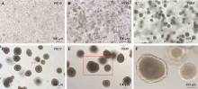
图1
小鼠食管消化后的单细胞在3D培养条件下形成囊状实心结构 A~E:小鼠食管组织被消化成单细胞后培养9天形成实心囊状物的过程;F:图E的局部放大图。"
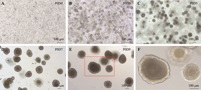
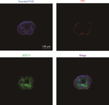
图2
食管类器官表达食管上皮细胞标志物P63和KRT13 培养至P1D9的食管类器官,使用抗体P63和KRT13进行免疫荧光染色结果。Hoechst33342试剂染细胞核,显示为蓝色;P63染基细胞细胞核,显示为红色;KRT13染上基层细胞细胞质,显示为绿色。"

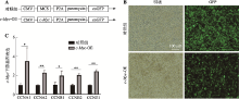
图3
成功构建表达外源基因的慢病毒 A:慢病毒质粒载体核心元件示意图。CMV为启动子,MCS表示多克隆位点(multiple cloning site)。B:慢病毒感染293T细胞72 h之后观察绿色荧光的结果。C:qPCR实验检测c-Myc下游基因表达情况。*:P<0.05;**:P<0.01。"
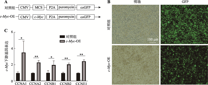

图4
食管类器官的慢病毒感染与筛选 慢病毒感染后的食管类器官在含2 μg/mL嘌呤霉素的培养基中培养至P1D7(即第1代第7天)的绿色荧光观察结果。"
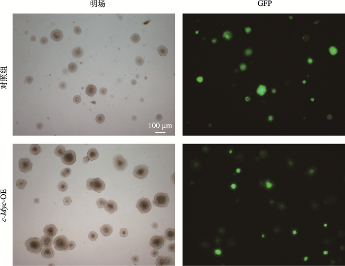

图5
qPCR和免疫荧光染色实验显示高效获得过表达c-Myc的食管类器官 A:感染后培养9天(P1D9)用qPCR检测总c-Myc的表达量;B:培养9天(P1D9)的食管类器官进行免疫荧光染色实验,用c-Myc抗体检测c-Myc的表达量。IF,Immunofluorescent staining method。***:P<0.001。"
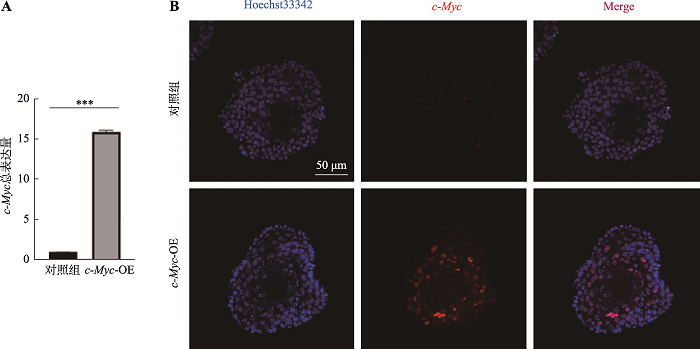
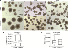
图6
c-Myc过表达对食管类器官的形态和大小均无显著影响 A:慢病毒感染后的类器官进行连续传代培养的过程,每代培养8~10天,所示为第一代、第四代、第7代培养9天后的生长情况;B:对照组和实验组类器官平均面积比较(P4D9);C:对照组和实验组类器官平均面积比较结果(P7D9)。ns:P>0.05,无显著差异。"

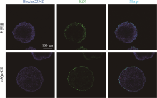
图7
免疫荧光染色显示Ki67的表达无显著变化 慢病毒感染后的食管类器官,用抗体Ki67进行免疫荧光染色的结果。"
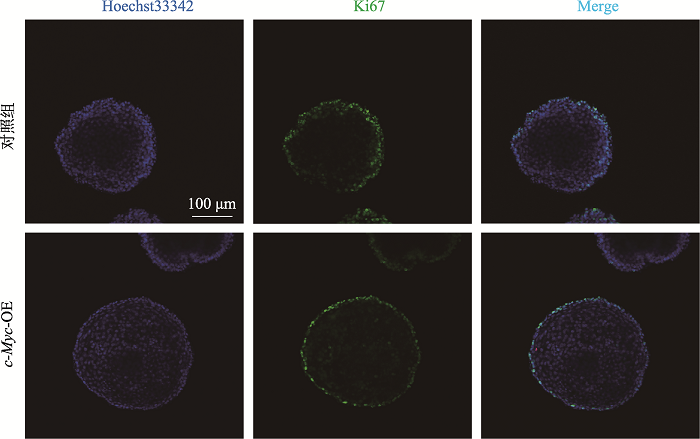
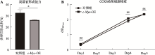
图8
食管类器官形成能力与用CCK8检测细胞增殖情况 A:相同细胞数培养形成的食管类器官数与对应细胞数的比例(食管类器官形成率);B:添加CCK8后测定吸光值(OD480 nm)。*:P<0.05;ns:P>0.05,无显著差异。"
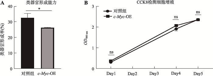
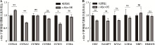
图9
q-PCR检测c-Myc下游与细胞周期、代谢相关的基因表达情况 A:c-Myc下游与细胞周期相关基因的表达量;B:c-Myc下游与细胞代谢相关基因的表达量。*:P<0.05;**:P<0.01;ns: P>0.05,无显著差异。"
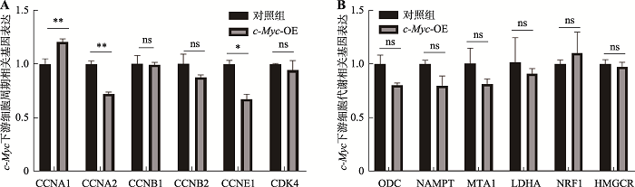
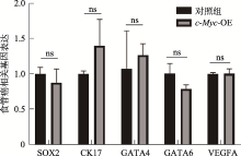
图10
食管癌相关基因的表达情况 qPCR检测食管癌相关基因表达情况。结果显示均无显著性差异(ns:P>0.05)。"
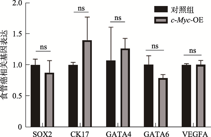
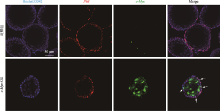
图1
附成熟食管类器官中c-Myc与P63共定位结果 用抗体c-Myc与P63对培养7天(Day7)的成熟食管类器官进行免疫荧光染色。图中白色箭头显示了同时表达c-Myc与P63的基底细胞。"
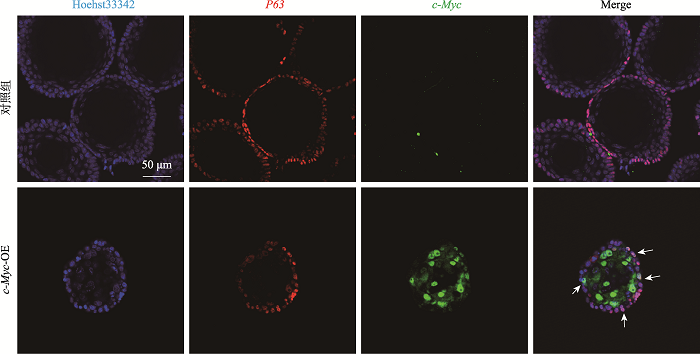

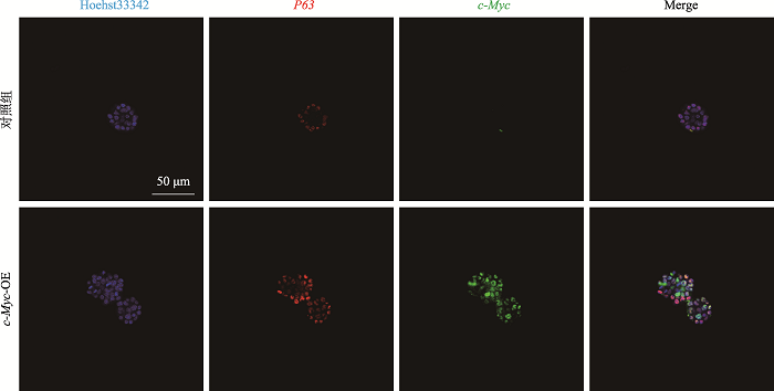
图2
附早期食管类器官中c-Myc与P63共定位结果 用抗体c-Myc与P63对培养7天(Day7)的早期食管类器官进行免疫荧光染色。大部分基底细胞均表达c-Myc与P63。"
| [1] | Lin YS, Totsuka Y, Shan BE, Wang CC, Wei WQ, Qiao YL, Kikuchi S, Inoue M, Tanaka H, He YT. Esophageal cancer in high-risk areas of China: research progress and challenges. Ann Epidemiol, 2017,27(3):215-221. |
| [2] | Huang FL, Yu SJ. Esophageal cancer: risk factors, genetic association, and treatment. Asian J Surg, 2018,41(3):210-215. |
| [3] | Sethi NS, Kikuchi O, Duronio GN, Stachler MD, McFarland JM, Ferrer-Luna R, Zhang YX, Bao CY, Bronson R, Patil D, Sanchez-Vega F, Liu JB, Sicinska E, Lazaro JB, Ligon KL, Beroukhim R, Bass AJ. Early TP53 alterations engage environmental exposures to promote gastric premalignancy in an integrative mouse model. Nat Genet, 2020,52(2):219-230. |
| [4] | Testa U, Castelli G, Pelosi E. Esophageal cancer: genomic and molecular characterization, stem cell compartment and clonal evolution. Medicines (Basel), 2017,4(3):67. |
| [5] | di Pietro M, Canto MI, Fitzgerald RC. Endoscopic management of early adenocarcinoma and squamous cell carcinoma of the esophagus: screening, diagnosis, and therapy. Gastroenterology, 2018,154(2):421-436. |
| [6] | Talukdar FR, di Pietro M, Secrier M, Moehler M, Goepfert K, Lima SSC, Pinto LFR, Hendricks D, Parker MI, Herceg Z. Molecular landscape of esophageal cancer: implications for early detection and personalized therapy. Ann N Y Acad Sci, 2018,1434(1):342-359. |
| [7] | Cancer Genome Atlas Research Network, Analysis Working Group: Asan University, BC Cancer Agency, Brigham and Women’s Hospital, Broad Institute, Brown University, Case Western Reserve University, Dana-Farber Cancer Institute, Duke University, Greater Poland Cancer Centre, Harvard Medical School, Institute for Systems Biology, KU Leuven, Mayo Clinic, Memorial Sloan Kettering Cancer Center, National Cancer Institute, Nationwide Children’s Hospital, Stanford University, University of Alabama, University of Michigan, University of North Carolina, University of Pittsburgh, University of Rochester, University of Southern California, University of Texas MD Anderson Cancer Center, University of Washington, Van Andel Research Institute, Vanderbilt University, Washington University, Genome Sequencing Center: Broad Institute, Washington University in St. Louis, Genome Characterization Centers: BC Cancer Agency, Broad Institute, Harvard Medical School, Sidney Kimmel Comprehensive Cancer Center at Johns Hopkins University, University of North Carolina, University of Southern California Epigenome Center, University of Texas MD Anderson Cancer Center, Van Andel Research Institute, Genome Data Analysis Centers: Broad Institute, Brown University, Harvard Medical School, Institute for Systems Biology, Memorial Sloan Kettering Cancer Center, University of California Santa Cruz, University of Texas MD Anderson Cancer Center, Biospecimen Core Resource: International Genomics Consortium, Research Institute at Nationwide Children’s Hospital, Tissue Source Sites: Analytic Biologic Services, Asan Medical Center, Asterand Bioscience, Barretos Cancer Hospital, BioreclamationIVT, Botkin Municipal Clinic, Chonnam National University Medical School, Christiana Care Health System, Cureline, Duke University, Emory University, Erasmus University, Indiana University School of Medicine, Institute of Oncology of Moldova, International Genomics Consortium, Invidumed, Israelitisches Krankenhaus Hamburg, Keimyung University School of Medicine, Memorial Sloan Kettering Cancer Center, National Cancer Center Goyang, Ontario Tumour Bank, Peter MacCallum Cancer Centre, Pusan National University Medical School, Ribeirão Preto Medical School, St. Joseph’s Hospital &Medical Center, St. Petersburg Academic University, Tayside Tissue Bank, University of Dundee, University of Kansas Medical Center, University of Michigan, University of North Carolina at Chapel Hill, University of Pittsburgh School of Medicine, University of Texas MD Anderson Cancer Center, Disease Working Group, Duke University, Memorial Sloan Kettering Cancer Center, National Cancer Institute, University of Texas MD Anderson Cancer Center, Yonsei University College of Medicine, Data Coordination Center: CSRA Inc, Project Team: National Institutes of Health. Integrated genomic characterization of oesophageal carcinoma. Nature, 2017,541(7636):169-175. |
| [8] | Lin DC, Dinh HQ, Xie JJ, Mayakonda A, Silva TC, Jiang YY, Ding LW, He JZ, Xu XE, Hao JJ, Wang MR, Li CQ, Xu LY, Li EM, Berman BP, Koeffler HP. Identification of distinct mutational patterns and new driver genes in oesophageal squamous cell carcinomas and adenocarcinomas. Gut, 2018,67(10):1769-1779. |
| [9] | Forbes SA, Beare D, Boutselakis H, Bamford S, Bindal N, Tate J, Cole CG, Ward S, Dawson E, Ponting L, Stefancsik R, Harsha B, Kok CY, Jia MM, Jubb H, Sondka Z, Thompson S, De T, Campbell PJ. COSMIC: somatic cancer genetics at high-resolution. Nucleic Acids Res, 2017,45(D1):D777-D783. |
| [10] | Hoffman B, Amanullah A, Shafarenko M, Liebermann DA. The proto-oncogene c-Myc in hematopoietic development and leukemogenesis. Oncogene, 2002,21(21):3414-3421. |
| [11] | Kim EY, Kim A, Kim SK, Chang YS. MYC expression correlates with PD-L1 expression in non-small cell lung cancer. Lung Cancer, 2017,110:63-67. |
| [12] | Dolezal JM, Wang HB, Kulkarni S, Jackson L, Lu J, Ranganathan S, Goetzman ES, Bharathi SS, Beezhold K, Byersdorfer CA, Prochownik EV. Sequential adaptive changes in a c-Myc-driven model of hepatocellular carcinoma. J Biol Chem, 2017,292(24):10068-10086. |
| [13] | Huang J, Jiang DX, Zhu T, Wang YQ, Wang H, Wang Q, Tan LJ, Zhu HG, Yao JX, Hou YY. Prognostic significance of c-Myc amplification in esophageal squamous cell carcinoma. Ann Thorac Surg, 2019,107(2):436-443. |
| [14] | von Rahden BHA, Stein HJ, Puhringer-Oppermann F, Sarbia M. c-Myc amplification is frequent in esophageal adenocarcinoma and correlated with the upregulation of VEGFA expression. Neoplasia, 2006,8(9):702-707. |
| [15] | Sun LL, Wang YQ, Cen J, Ma XL, Cui L, Qiu ZX, Zhang ZT, Li H, Yang RZ, Wang CH, Chen XT, Wang L, Ye Y, Zhang HB, Pan GY, Kang JS, Ji Y, Zheng YW, Zheng S, Hui LJ. Modelling liver cancer initiation with organoids derived from directly reprogrammed human hepatocytes. Nat Cell Biol, 2019,21(8):1015-1026. |
| [16] | Clevers H. Modeling development and disease with organoids. Cell, 2016,165(7):1586-1597. |
| [17] | Huch M, Koo BK. Modeling mouse and human development using organoid cultures. Development, 2015,142(18):3113-3125. |
| [18] | Schutgens F, Clevers H. Human organoids: tools for understanding biology and treating diseases. Annu Rev Pathol, 2020,15:211-234. |
| [19] | Trisno SL, Philo KED, McCracken KW, Catá EM, Ruiz-Torres S, Rankin SA, Han L, Nasr T, Chaturvedi P, Rothenberg ME, Mandegar MA, Wells SI, Zorn AM, Wells JM,. Esophageal organoids from human pluripotent stem cells delineate SOX2 functions during esophageal specification. Cell Stem Cell, 2018, 23(4): 501-515.e7. |
| [20] | Zhang YC, Yang Y, Jiang M, Huang SX, Zhang WW, Alam DA, Danopoulos S, Mori M, Chen YW, Balasubramanian R, de Sousa Lopes SMC, Serra C, Bialecka M, Kim E, Lin SJ, de Carvalho ALRT, Riccio PN, Cardoso WV, Zhang X, Snoeck HW, Que JW. 3D Modeling of esophageal development using human PSC-derived basal progenitors reveals a critical role for notch signaling. Cell Stem Cell, 2018, 23(4): 516-529.e5. |
| [21] | Bailey DD, Zhang YC, van Soldt BJ, Jiang M, Suresh S, Nakagawa H, Rustgi AK, Aceves SS, Cardoso WV, Que JW. Use of hPSC-derived 3D organoids and mouse genetics to define the roles of YAP in the development of the esophagus. Development, 2019, 146(23): xdev178855. |
| [22] | Fujii M, Clevers H, Sato T. Modeling human digestive diseases with CRISPR-Cas9-modified organoids. Gastroenterology, 2019,156(3):562-576. |
| [23] | DeWard AD, Cramer J, Lagasse E. Cellular heterogeneity in the mouse esophagus implicates the presence of a nonquiescent epithelial stem cell population. Cell Rep, 2014,9(2):701-711. |
| [24] | Koo BK, Sasselli V, Clevers H. Retroviral gene expression control in primary organoid cultures. Curr Protoc Stem Cell Biol, 2013, 27: Unit 5A.6. |
| [25] | Maru Y, Orihashi K, Hippo Y. Lentivirus-based stable gene delivery into intestinal organoids. Methods Mol Biol, 2016,1422:13-21. |
| [26] | Wei JS, Ran G, Wang X, Jiang N, Liang JQ, Lin XH, Ling C, Zhao B. Gene manipulation in liver ductal organoids by optimized recombinant adeno-associated virus vectors. J Biol Chem, 2019,294(38):14096-14104. |
| [27] | Sachs N, Papaspyropoulos A, Zomer-van Ommen DD, Heo I, Böttinger L, Klay D, Weeber F, Huelsz-Prince G, Iakobachvili N, Amatngalim GD, de Ligt J, van Hoeck A, Proost N, Viveen MC, Lyubimova A, Teeven L, Derakhshan S, Korving J, Begthel H, Dekkers JF, Kumawat K, Ramos E, van Oosterhout MF, Offerhaus GJ, Wiener DJ, Olimpio EP, Dijkstra KK, Smit EF, van der Linden M, Jaksani S, van de Ven M, Jonkers J, Rios AC, Voest EE, van Moorsel CH, van der Ent CK, Cuppen E, van Oudenaarden A, Coenjaerts FE, Meyaard L, Bont LJ, Peters PJ, Tans SJ, van Zon JS, Boj SF, Vries RG, Beekman JM, Clevers H. Long-term expanding human airway organoids for disease modeling. EMBO J, 2019,38(4):e100300. |
| [28] | Dang CV. Myc on the path to cancer. Cell, 2012,149(1):22-35. |
| [29] | Dang CV. Myc, metabolism, cell growth, and tumorigenesis. Cold Spring Harb Perspect Med, 2013,3(8):a014217. |
| [30] | Li J, Liang Y, Lv H, Meng H, Xiong G, Guan XY, Chen XD, Bai Y, Wang K. miR-26a and miR-26b inhibit esophageal squamous cancer cell proliferation through suppression of c-Myc pathway. Gene, 2017,625:1-9. |
| [31] | McCauley HA, Wells JM. Pluripotent stem cell-derived organoids: using principles of developmental biology to grow human tissues in a dish. Development, 2017,144(6):958-962. |
| [32] | Pan XN, Chen JJ, Wang LX, Xiao RZ, Liu LL, Fang ZG, Liu QT, Long ZJ, Lin DJ. Inhibition of c-Myc overcomes cytotoxic drug resistance in acute myeloid leukemia cells by promoting differentiation. PLoS One, 2014,9(8):e105381. |
| [33] | Chan JC, Hannan KM, Riddell M, Ng PY, Peck A, Lee RS, Hung S, Astle MV, Bywater M, Wall M, Poortinga G, Jastrzebski K, Sheppard KE, Hemmings BA, Hall MN, Johnstone RW, McArthur GA, Hannan RD, Pearson RB. AKT promotes rRNA synthesis and cooperates with c-Myc to stimulate ribosome biogenesis in cancer. Sci Signal, 2011, 4(188): ra56. |
| [34] | Liang MQ, Yu FQ, Chen C. C-Myc regulates PD-L1 expression in esophageal squamous cell carcinoma. Am J Transl Res, 2020,12(2):379-388. |
| [35] | Hong J, Maacha S, Belkhiri A. Transcriptional upregulation of c-Myc by AXL confers epirubicin resistance in esophageal adenocarcinoma. Mol Oncol, 2018,12(12):2191-2208. |
| [36] | Rygiel AM, Milano F, Ten Kate FJ, Schaap A, Wang KK, Peppelenbosch MP, Bergman JJGHM, Krishnadath KK. Gains and amplifications of c-Myc, EGFR, and 20.q13 loci in the no dysplasia-dysplasia-adenocarcinoma sequence of Barrett's esophagus. Cancer Epidemiol Biomarkers Prev, 2008,17(6):1380-1385. |
| [1] | 史悦,许争争,鲁欢,慈维敏. 肿瘤突变特征与病理分型的关联研究[J]. 遗传, 2018, 40(11): 1033-1038. |
| [2] | 郝佳洁, 王春丽, 顾文跃, 程潇钰, 张钰, 徐昕, 蔡岩, 王明荣. 利用染色体区段混合BAC探针鉴定食管癌细胞中的染色体畸变[J]. 遗传, 2014, 36(6): 558-565. |
| [3] | 杨扬,王博石,汪晓敏,张钰,王明荣,贾雪梅. 食管癌细胞抗失巢凋亡基因UBCH7的发现与鉴定[J]. 遗传, 2012, 34(2): 190-197. |
| [4] | 韩亚玲,冯彦斌,罗曼莉,徐昕,蔡岩,王明荣. 人食管癌EC9706单克隆的分离培养及其生物学特性的研究[J]. 遗传, 2007, 29(11): 1331-1335. |
| [5] | 龚嫦虹,邱定红,黄鹰,周晓雷,王为未,张丽珊. P16基因与散发性食管癌的研究[J]. 遗传, 2000, 22(1): 7-10. |
| 阅读次数 | ||||||
|
全文 |
|
|||||
|
摘要 |
|
|||||

www.chinagene.cn
备案号:京ICP备09063187号-4
总访问:,今日访问:,当前在线:
