
遗传 ›› 2025, Vol. 47 ›› Issue (3): 382-392.doi: 10.16288/j.yczz.24-213
• 技术与方法 • 上一篇
高炳熙( ), 吴华煊, 杜志强()
收稿日期:2024-07-19
修回日期:2024-09-23
出版日期:2025-03-20
发布日期:2024-09-26
通讯作者:
杜志强,博士,教授,研究方向:动物遗传育种与繁殖。E-mail: zhqdu@yangtzeu.edu.cn作者简介:高炳熙,硕士研究生,专业方向:动物遗传育种与繁殖。E-mail: gbx15020771250@163.com
基金资助:
Bingxi Gao(), Huaxuan Wu, Zhiqiang Du()
Received:2024-07-19
Revised:2024-09-23
Published:2025-03-20
Online:2024-09-26
Supported by:摘要:
单细胞转录组测序(single-cell transcriptome sequencing, scRNA-seq)通过高通量获取单细胞转录丰度数据,能够深入揭示细胞类型、亚型组成、特异性基因标记及功能差异,广泛应用于动植物发育生物学和重要性状解析等领域。然而,scRNA-seq数据常伴随高噪声、高维度和批次效应等问题,导致大量低表达基因和变异的出现,严重影响数据分析的准确性和可靠性。这不仅增加了数据处理的复杂性,还制约了特征选择和下游分析的效果。尽管已有多种统计推断和机器学习方法用于应对这些挑战,但在细胞类型识别、特征选择以及批次效应校正等方面,现有方法仍存在着局限性,难以满足复杂生物学研究的需求。因此本研究提出了一种创新的单细胞分类方法scIC (single-cell image classification),将scRNA-seq数据转换为图像形式,并结合深度学习技术进行细胞分类。通过这种图像转换的方式能够更有效地捕捉数据中的复杂模式,进而利用卷积神经网络(convolutional neural networks, CNN)和残差网络(residual network, ResNet)构建高效的分类模型。在测试4种细胞类型(小鼠皮肤基底细胞、小鼠淋巴细胞、人类神经元细胞和小鼠脊髓细胞)的scRNA-seq数据后,分类模型的准确率均超过94%,其中小鼠皮肤基底细胞数据集使用ResNet50模型时的分类准确率高达99.8%。这些结果表明,将scRNA-seq数据进行图像转换并与深度学习技术结合,能够显著提高分类准确性,为解决单细胞数据分析中的关键挑战提供了新思路和有效工具。本研究代码已公开于:
高炳熙, 吴华煊, 杜志强. 应用图像转换与深度学习提升单细胞分类精度[J]. 遗传, 2025, 47(3): 382-392.
Bingxi Gao, Huaxuan Wu, Zhiqiang Du. Enhancing single-cell classification accuracy using image conversion and deep learning[J]. Hereditas(Beijing), 2025, 47(3): 382-392.
表1
本研究使用的数据来源"
| 数据集 | 样本数 | 基因数 | 类别数 |
|---|---|---|---|
| 小鼠皮肤基底细胞 | 9,288 | 1,222 | 3 |
| 小鼠淋巴细胞 | 11,127 | 14,918 | 6 |
| 人类神经元细胞 | 306 | 27,760 | 8 |
| 小鼠脊髓细胞 | 22,155 | 18,772 | 10 |
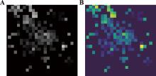
图1
基因表达矩阵可视化 A:灰度图;B:热图。"
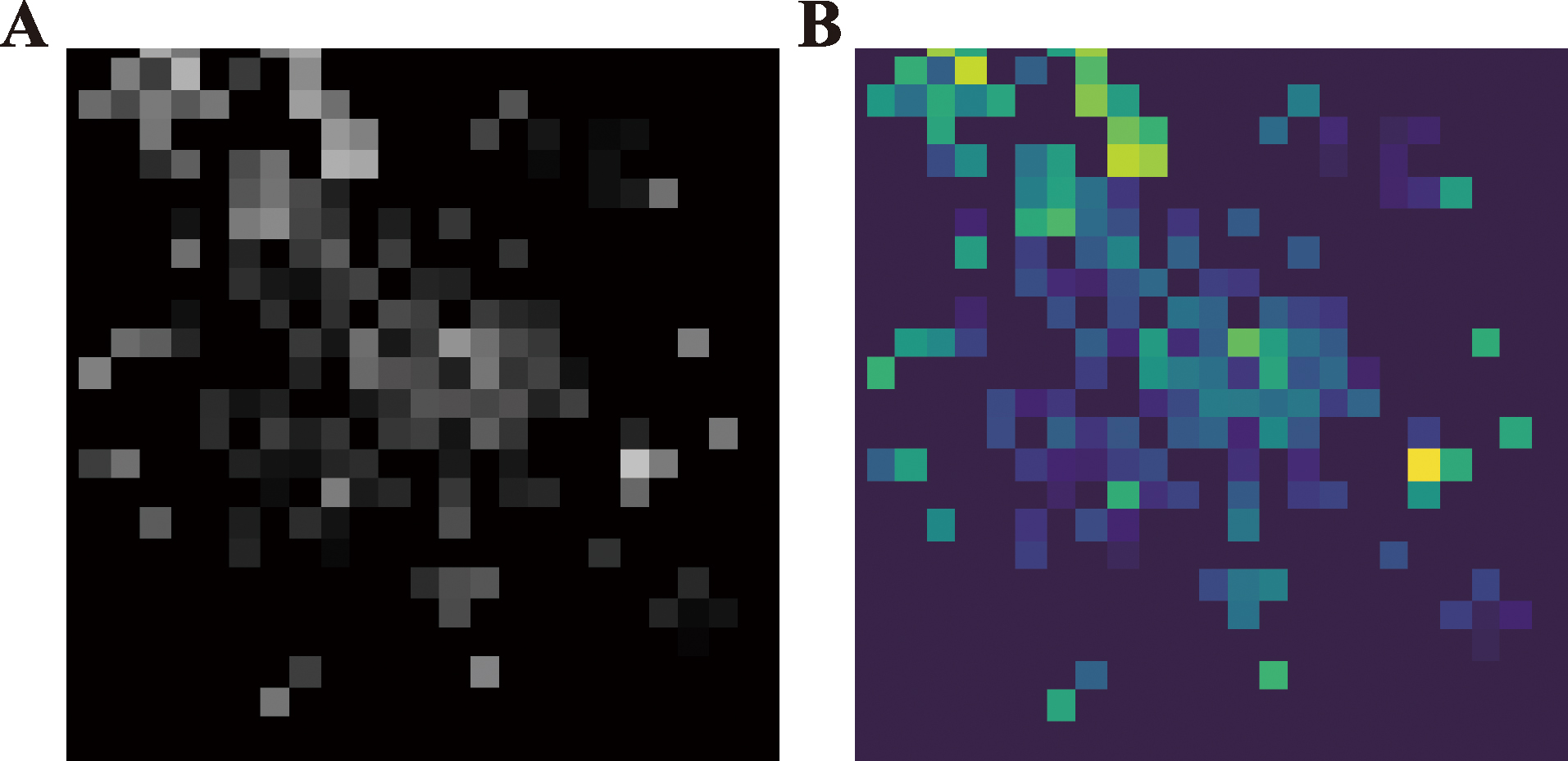
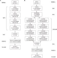
图2
分类模型结构可视化"

表2
7种聚类方法性能指标"
| 数据集 | 指标 | 聚类方法 | |||||||
|---|---|---|---|---|---|---|---|---|---|
| Grayscale image | Heat map | scPoli | scDML | AHC | GMM | BRICH | SC | ||
| 小鼠皮肤基底细胞 | ARI | 0.988 | 0.993 | 0.989 | 0.987 | 0.984 | 0.992 | 0.985 | 0.751 |
| NMI | 0.972 | 0.982 | 0.982 | 0.971 | 0.962 | 0.982 | 0.967 | 0.743 | |
| 小鼠淋巴细胞 | ARI | 0.752 | 0.856 | 0.793 | 0.714 | 0.842 | 0.661 | 0.662 | 0.58 |
| NMI | 0.76 | 0.79 | 0.776 | 0.740 | 0.825 | 0.761 | 0.735 | 0.76 | |
| 人类神经元细胞 | ARI | 0.848 | 0.816 | 0.825 | 0.697 | 0.663 | 0.642 | 0.713 | 0.62 |
| NMI | 0.876 | 0.845 | 0.843 | 0.803 | 0.732 | 0.743 | 0.782 | 0.71 | |
| 小鼠脊髓细胞 | ARI | 0.715 | 0.723 | 0.677 | 0.524 | 0.164 | 0.447 | 0.168 | 0.01 |
| NMI | 0.763 | 0.781 | 0.695 | 0.723 | 0.399 | 0.241 | 0.392 | 0.01 | |
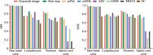
图3
不同聚类方法ARI和NMI指标 A:不同模型ARI评分;B:不同模型NMI评分。"

表3
细胞分类模型评价指标"
| 数据集 | 指标 | 细胞分类模型 | |||||
|---|---|---|---|---|---|---|---|
| SnapCCESS | scMDC | CNN | ResNet18 | ResNet34 | ResNet50 | ||
| 小鼠皮肤基底细胞 | ACC | 0.995(0.002) | 0.994(0.003) | 0.995 (0.002) | 0.995 (0.002) | 0.994 (0.003) | 0.998 (0.001) |
| P | 0.995(0.003) | 0.995(0.003) | 0.995 (0.002) | 0.995 (0.002) | 0.994 (0.002) | 0.997 (0.001) | |
| R | 0.995(0.003) | 0.996(0.003) | 0.995 (0.003) | 0.995 (0.003) | 0.994 (0.003) | 0.998 (0.002) | |
| F1 | 0.994(0.002) | 0.996(0.002) | 0.995 (0.002) | 0.995 (0.003) | 0.994 (0.003) | 0.998 (0.001) | |
| Loss | 0.040 (0.005) | 0.020(0.005) | 0.030 (0.005) | 0.010 (0.005) | 0.020 (0.006) | 0.010 (0.003) | |
| 小鼠淋巴细胞 | ACC | 0.935(0.006) | 0.935(0.005) | 0.934 (0.005) | 0.941 (0.003) | 0.938 (0.004) | 0.934 (0.005) |
| P | 0.934(0.002) | 0.937(0.004) | 0.907 (0.004) | 0.929 (0.003) | 0.912 (0.003) | 0.920 (0.004) | |
| R | 0.932(0.005) | 0.936(0.004) | 0.856 (0.006) | 0.900 (0.004) | 0.896 (0.004) | 0.891 (0.006) | |
| F1 | 0.941(0.005) | 0.937(0.004) | 0.874 (0.005) | 0.905 (0.003) | 0.902 (0.004) | 0.896 (0.005) | |
| Loss | 0.310 (0.005) | 0.230(0.005) | 0.210 (0.010) | 0.190 (0.006) | 0.220 (0.008) | 0.210 (0.001) | |
| 人类神经元细胞 | ACC | 0.933(0.029) | 0.926(0.026) | 0.967 (0.015) | 0.961 (0.018) | 0.925 (0.025) | 0.849 (0.031) |
| P | 0.937(0.031) | 0.932(0.023) | 0.963 (0.012) | 0.959 (0.014) | 0.932 (0.021) | 0.819 (0.028) | |
| R | 0.947(0.027) | 0.947(0.030) | 0.958 (0.018) | 0.945 (0.021) | 0.891 (0.030) | 0.787 (0.042) | |
| F1 | 0.947(0.023) | 0.947(0.030) | 0.953 (0.015) | 0.946 (0.018) | 0.890 (0.025) | 0.776 (0.035) | |
| Loss | 0.130 (0.051) | 0.142(0.042) | 0.320 (0.030) | 0.150 (0.036) | 0.390 (0.050) | 0.600 (0.070) | |
| 小鼠脊髓细胞 | ACC | 0.952(0.007) | 0.957(0.003) | 0.944 (0.008) | 0.958 (0.008) | 0.960 (0.007) | 0.958 (0.009) |
| P | 0.942(0.003) | 0.945(0.005) | 0.855 (0.008) | 0.959 (0.006) | 0.961 (0.006) | 0.957 (0.007) | |
| R | 0.932(0.007) | 0.928(0.012) | 0.835 (0.012) | 0.912 (0.009) | 0.939 (0.008) | 0.934 (0.009) | |
| F1 | 0.925(0.008) | 0.934(0.013) | 0.844 (0.010) | 0.929 (0.008) | 0.947 (0.007) | 0.942 (0.009) | |
| Loss | 0.130 (0.005) | 0.210(0.006) | 0.200 (0.020) | 0.130 (0.016) | 0.130 (0.014) | 0.140 (0.018) | |
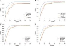
图4
不同模型训练过程可视化 A:CNN模型;B:ResNet18 模型;C:ResNet34模型;D:ResNet50模型。"
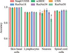
图5
模型准确率对比"
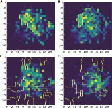
图6
细胞分类模型LIME分析可视化 A:CNN模型;B:ResNet18模型;C:ResNet34模型;D:ResNet50模型。"

| [1] |
Giladi A, Amit I. Single-cell genomics: a stepping stone for future immunology discoveries. Cell, 2018, 172(1-2): 14-21.
doi: S0092-8674(17)31320-X pmid: 29328909 |
| [2] | Han H, Luo FC. Application of single-cell RNA sequencing in probing oligodendroglia heterogeneity and neurological disorders. Hereditas (Beijing), 2023, 45(3): 198-211. |
| 韩熙, 罗富成. 单细胞转录组测序在少突胶质谱系细胞异质性与神经系统疾病中的应用. 遗传, 2023, 45(3): 198-211. | |
| [3] | Qu L, Li S, Qiu HJ. Applications of single-cell RNA sequencing in virology. Hereditas (Beijing), 2020, 42(3): 269-277. |
| 屈亮, 李素, 仇华吉. 单细胞RNA测序技术在病毒研究中的应用. 遗传, 2020, 42(3): 269-277. | |
| [4] | Zhang CQ, Geng Y, Han ZB, Liu YQ, Fu HZ, Hu QH. Autoencoder in autoencoder networks. IEEE Trans Neural Netw Learn Syst, 2024, 35(2): 2263-2275. |
| [5] | Li ZW, Liu F, Yang WJ, Peng SH, Zhou J. A survey of convolutional neural networks: analysis, applications, and prospects. IEEE Trans Neural Netw Learn Syst, 2022, 33(12): 6999-7019. |
| [6] | Arafa A, El-Fishawy N, Badawy M, Radad M. RNAutoencoder: reduced noise autoencoder for classifying imbalanced cancer genomic data. J Biol Eng, 2023, 17(1): 7. |
| [7] | Zhao JP, Wang N, Wang HY, Zheng CH, Su YS. SCDRHA: a scrna-seq data dimensionality reduction algorithm based on hierarchical autoencoder. Front Genet, 2021, 12: 733906. |
| [8] | Zhu YJ, Chen FX. Research progress of object recognition methods based on machine vision. Sci. Technol. Soc. 2023, 21(21): 21-24. |
| 朱亚军, 陈砆兴. 基于机器视觉的目标识别方法的研究进展. 科技资讯, 2023, 21(21): 21-24. | |
| [9] | Eltager M, Abdelaal T, Mahfouz A, Reinders MJT. scMoC: single-cell multi-omics clustering. Bioinform Adv, 2022, 2(1): vbac011. |
| [10] |
Song Q, Wang JT, Bar-Joseph Z. scSTEM: clustering pseudotime ordered single-cell data. Genome Biol, 2022, 23(1): 150.
doi: 10.1186/s13059-022-02716-9 pmid: 35799304 |
| [11] |
Islam MT, Xing L. Cartography of genomic interactions enables deep analysis of single-cell expression data. Nat Commun, 2023, 14(1): 679.
doi: 10.1038/s41467-023-36383-6 pmid: 36755047 |
| [12] | Jia SR, Lysenko A, Boroevich KA, Sharma A, Tsunoda T. scDeepInsight: a supervised cell-type identification method for scRNA-seq data with deep learning. Brief Bioinform, 2023, 24(5): bbad266. |
| [13] | Du ZH, Hu WL, Li JQ, Shang XQ, You ZH, Chen ZZ, Huang YA. scPML: pathway-based multi-view learning for cell type annotation from single-cell RNA-seq data. Commun Biol, 2023, 6(1): 1268. |
| [14] |
Derry A, Krzywinski M, Altman N. Convolutional neural networks. Nat Methods, 2023, 20(9): 1269-1270.
doi: 10.1038/s41592-023-01973-1 pmid: 37580560 |
| [15] | He FX, Liu TL, Tao DC. Why resnet works? Residuals generalize. IEEE Trans Neural Netw Learn Syst, 2020, 31(12): 5349-5362. |
| [16] |
Yu XK, Xu XY, Zhang JX, Li XJ. Batch alignment of single-cell transcriptomics data using deep metric learning. Nat Commun, 2023, 14(1): 960.
doi: 10.1038/s41467-023-36635-5 pmid: 36810607 |
| [17] |
Sharma A, Vans E, Shigemizu D, Boroevich KA, Tsunoda T. DeepInsight: a methodology to transform a non-image data to an image for convolution neural network architecture. Sci Rep, 2019, 9(1): 11399.
doi: 10.1038/s41598-019-47765-6 pmid: 31388036 |
| [18] |
Kang MJ, Lee S, Lee D, Kim S. Learning cell-type-specific gene regulation mechanisms by multi-attention based deep learning with regulatory latent space. Front Genet, 2020, 11: 869.
doi: 10.3389/fgene.2020.00869 pmid: 33133123 |
| [19] | Bao YC, Shi CX, Zhang CQ, Gu MJ, Zhu L, Liu ZX, Zhou L, Ma FY, Na RS, Zhang WG. Progress on deep learning in genomics. Hereditas (Beijing), 2024, 46(9): 701-715. |
| 鲍艳春, 石彩霞, 张传强, 谷明娟, 朱琳, 刘在霞, 周乐, 马凤英, 娜日苏, 张文广. 深度学习在基因组学中的研究进展. 遗传, 2024, 46(9): 701-715. | |
| [20] | Zheng HY, Wu HX, Du ZQ. Gut macrogenomic image enhancement and deep learning improve metabolic disease classification prediction accuracy. Hereditas (Beijing), 2024, 46(10): 886-896. |
| 郑慧怡, 吴华煊, 杜志强. 肠道宏基因组图像增强和深度学习改善代谢性疾病分类预测精度. 遗传, 2024, 46(10): 886-896. | |
| [21] |
Padovani F, Mairhörmann B, Falter-Braun P, Lengefeld J, Schmoller KM. Segmentation, tracking and cell cycle analysis of live-cell imaging data with Cell-ACDC. BMC Biol, 2022, 20(1): 174.
doi: 10.1186/s12915-022-01372-6 pmid: 35932043 |
| [22] | Salvatore M, Horlacher M, Marsico A, Winther O, Andersson R. Transfer learning identifies sequence determinants of cell-type specific regulatory element accessibility. NAR Genom Bioinform, 2023, 5(2): lqad026. |
| [23] | Lou RD, Chen JB, Hou HH, Liu YL, Tian Z, Zhang PC, Gui ZG. A new method of cell classification based on deep convolution neural networks. Journal of Test and Measurement Technology, 2019, 33(6): 509-515. |
| 娄润东, 陈俊彪, 侯宏花, 刘艳莉, 田珠, 张鹏程, 桂志国. 基于深度卷积神经网络的细胞分类新方法. 测试技术学报, 2019, 33(6): 509-515. | |
| [24] | Alaeddine H, Jihene M. Deep residual network in network. Comput Intell Neurosci, 2021, 2021: 6659083. |
| [25] |
De Donno C, Hediyeh-Zadeh S, Moinfar AA, Wagenstetter M, Zappia L, Lotfollahi M, Theis FJ. Population-level integration of single-cell datasets enables multi-scale analysis across samples. Nat Methods, 2023, 20(11): 1683-1692.
doi: 10.1038/s41592-023-02035-2 pmid: 37813989 |
| [26] | Yu LJ, Liu CL, Yang JYH, Yang PY. Ensemble deep learning of embeddings for clustering multimodal single- cell omics data. Bioinformatics, 2023, 39(6): btad382. |
| [27] |
Lin X, Tian T, Wei Z, Hakonarson H. Clustering of single-cell multi-omics data with a multimodal deep learning method. Nat Commun, 2022, 13(1): 7705.
doi: 10.1038/s41467-022-35031-9 pmid: 36513636 |
| [28] | Amin MR, Hasan M, Arnab SP, DeGiorgio M. Tensor decomposition-based feature extraction and classification to detect natural selection from genomic data. Mol Biol Evol. 2023 Oct 4; 40(10): msad216. |
| [29] |
Wu TC, Yu JX, Huang XF, Chen S, Wang YX, Pu YM. Preliminary study on deep learning picture classification model for identification and classification of invasion pattern of oral squamous cell carcinoma. J Oral Sci Res, 2023, 39(10): 917-922.
doi: 10.13701/j.cnki.kqyxyj.2023.10.013 |
|
吴天赐, 郁佳鑫, 黄晓峰, 陈盛, 王育新, 蒲玉梅. 深度学习图片分类模型ResNet-18用于判定口腔鳞状细胞癌浸润方式的初步研究. 口腔医学研究, 2023, 39(10): 917-922.
doi: 10.13701/j.cnki.kqyxyj.2023.10.013 |
|
| [30] | Qiao L, Wu WN, Lu ZT. Radiomics-based cerebrospinal fluid cell classification. Chinese Journal of Medical Physics, 2023, 40(2): 244-250. |
| 乔琳, 吴文娜, 卢振泰. 基于影像组学的脑脊液细胞分类方法. 中国医学物理学杂志, 2023, 40(2): 244-250. | |
| [31] | Liu M, Zhou L. Classification of cervical cells based on transfer learning and label smoothing strategy. Modern Computer, 2022, 28(19): 1-9+32. |
| 刘美, 周龙. 基于迁移学习与标签平滑策略的宫颈细胞分类方法. 现代计算机, 2022, 28(19): 1-9+32. | |
| [32] | Wu FQ, Lv LL, Lv D, Feng CB, Shi T, Wang W, Cui HH, Zhou Y. Deep learning model for automatic recognition of erythroid cells and granulocyte cells in bone marrow. Journal of Jilin University(lnformation Science Edition). 2020, 38(6): 729-736. |
| 吴汾奇, 吕丽丽, 吕迪, 冯辰彬, 施恬, 王维, 崔红花, 周柚. 骨髓红粒细胞自动识别的深度学习模型. 吉林大学学报(信息科学版), 2020, 38(6): 729-736. | |
| [33] | Sun K, Yao XF, Ma FL, Zhao WS, Huang G. Blood cell classification based on machine learning. Chinese Journal of Medical Physics, 2020, 37(1): 127-132. |
| 孙凯, 姚旭峰, 马风玲, 赵文硕, 黄钢. 基于机器学习的血细胞分类研究进展. 中国医学物理学杂志, 2020, 37(01): 127-132. | |
| [34] |
Frank SM, Qi A, Ravasio D, Sasaki Y, Rosen EL, Watanabe T. Supervised learning occurs in visual perceptual learning of complex natural images. Curr Biol, 2020, 30(15): 2995-3000.e3.
doi: S0960-9822(20)30737-5 pmid: 32502415 |
| [35] | Ju W, Luo X, Ma ZY, Yang JW, Deng MH, Zhang M. GHNN: graph harmonic neural networks for semi-supervised graph-level classification. Neural Netw, 2022, 151: 70-79. |
| [36] |
Kiselev VY, Andrews TS, Hemberg M. Challenges in unsupervised clustering of single-cell rna-seq data. Nat Rev Genet, 2019, 20(5): 273-282.
doi: 10.1038/s41576-018-0088-9 pmid: 30617341 |
| [37] | Stahlschmidt SR, Ulfenborg B, Synnergren J. Multimodal deep learning for biomedical data fusion: a review. Brief Bioinform, 2022, 23(2): bbab569. |
| [38] | Felix MA, Wagner A. Robustness and evolution: concepts, insights and challenges from a developmental model system. Heredity (Edinb), 2008, 100(2): 132-140. |
| [1] | 鲍艳春, 石彩霞, 张传强, 谷明娟, 朱琳, 刘在霞, 周乐, 马凤英, 娜日苏, 张文广. 深度学习在基因组学中的研究进展[J]. 遗传, 2024, 46(9): 701-715. |
| [2] | 杨帆, 韩巧玲, 赵文迪, 赵玥. 基于层级和全局特征结合的蛋白质序列EC编号预测[J]. 遗传, 2024, 46(8): 661-669. |
| [3] | 郑慧怡, 吴华煊, 杜志强. 肠道宏基因组图像增强和深度学习改善代谢性疾病分类预测精度[J]. 遗传, 2024, 46(10): 886-896. |
| [4] | 周俊, 赵成成, 吴霄, 石俊松, 周荣, 吴珍芳, 李紫聪. 猪耳成纤维细胞转录组异质性及对核移植胚胎发育的潜在影响[J]. 遗传, 2020, 42(9): 898-915. |
| [5] | 张强, 顾明亮. 单细胞测序技术及其在乳腺癌研究中的应用[J]. 遗传, 2020, 42(3): 250-268. |
| [6] | 吴保军,王卓,董宇,邓宇亮,施奇惠. 肺癌恶性胸腔积液中稀有肿瘤细胞的鉴定与单细胞测序分析[J]. 遗传, 2019, 41(2): 175-184. |
| [7] | 胡伟澎, 李佑平, 张秀清. 基于迁移学习的MHC-I型抗原表位呈递预测[J]. 遗传, 2019, 41(11): 1041-1049. |
| [8] | 康岚, 陈嘉瑜, 高绍荣. 中国细胞重编程和多能干细胞研究进展[J]. 遗传, 2018, 40(10): 825-840. |
| 阅读次数 | ||||||
|
全文 |
|
|||||
|
摘要 |
|
|||||

www.chinagene.cn
备案号:京ICP备09063187号-4
总访问:,今日访问:,当前在线:
