
遗传 ›› 2023, Vol. 45 ›› Issue (3): 212-220.doi: 10.16288/j.yczz.22-422
商晓康( ), 张思萌(), 倪军军()
收稿日期:2022-12-26
修回日期:2023-02-15
出版日期:2023-03-20
发布日期:2023-02-21
通讯作者:
倪军军
E-mail:1120202339@bit.edu.cn;zhangsimeng0301@163.com;nijunjun@bit.edu.cn
作者简介:商晓康,在读本科生,专业方向:生物技术。E-mail: 基金资助:
Xiaokang Shang(), Simeng Zhang(), Junjun Ni()
Received:2022-12-26
Revised:2023-02-15
Online:2023-03-20
Published:2023-02-21
Contact:
Ni Junjun
E-mail:1120202339@bit.edu.cn;zhangsimeng0301@163.com;nijunjun@bit.edu.cn
Supported by:摘要:
组织蛋白酶B (cathepsin B,CatB)是一种定位于溶酶体的半胱氨酸蛋白酶,最初被认为在溶酶体内发挥非选择性地降解吞噬或者自噬蛋白的功能。然而最新研究发现,CatB也可以选择性地降解或特异性地活化目标蛋白,从而参与调控生理病理反应。在衰老及相关的神经退行性疾病的大脑中,CatB的表达、酶活性及细胞定位都发生了显著变化,因此CatB在衰老和神经退行性疾病中的病理学功能备受关注。本文对CatB参与脑衰老及阿尔兹海默症进程的相关研究进行了系统梳理,并讨论了目前有关CatB的神经病理学研究中存在的问题,以期为全面认识脑衰老及阿尔兹海默症的病理机制奠定基础。
商晓康, 张思萌, 倪军军. 组织蛋白酶B参与脑衰老及阿尔兹海默症发生发展研究进展[J]. 遗传, 2023, 45(3): 212-220.
Xiaokang Shang, Simeng Zhang, Junjun Ni. Research progress of cathepsin B in brain aging and Alzheimer’s diseases[J]. Hereditas(Beijing), 2023, 45(3): 212-220.
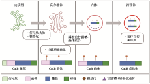
图1
CatB在细胞器中的转移及成熟过程 在内质网中,CatB酶原被切除信号肽且被糖基化;随后转移到高尔基体,CatB前体的甘露糖残基被磷酸化后,与内体中甘露糖-6磷酸化受体结合,并进一步在溶酶体酸性环境中发生前体自切割,形成CatB成熟体。"
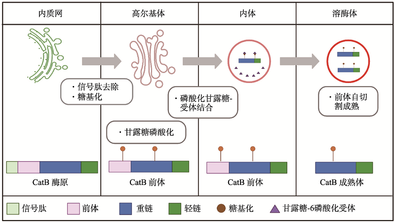
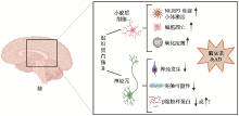
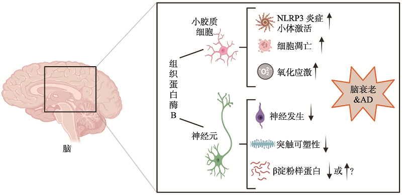
图2
CatB在脑衰老及AD病理中的功能 在大脑中,CatB分别在小胶质细胞和神经元中表达。在小胶质细胞中,CatB介导NLRP3炎症小体激活、细胞凋亡和氧化应激;在神经元中,CatB会破坏神经发生和突触可塑性,并且对β淀粉样蛋白有影响,但是CatB对β淀粉样蛋白诱导或清除的研究还未形成统一的结论。"
| [1] | Ni JJ, Lan F, Xu Y, Nakanishi H, Li X. Extralysosomal cathepsin B in central nervous system: mechanisms and therapeutic implications. Brain Pathol, 2022, 32(5): e13071. |
| [2] |
Musil D, Zucic D, Turk D, Engh RA, Mayr I, Huber R, Popovic T, Turk V, Towatari T, Katunuma N. The refined 2.15 A X-ray crystal structure of human liver cathepsin B: the structural basis for its specificity. EMBO J, 1991, 10(9): 2321-2330.
doi: 10.1002/j.1460-2075.1991.tb07771.x pmid: 1868826 |
| [3] |
Illy C, Quraishi O, Wang J, Purisima E, Vernet T, Mort JS. Role of the occluding loop in cathepsin B activity. J Biol Chem, 1997, 272(2): 1197-1202.
doi: 10.1074/jbc.272.2.1197 pmid: 8995421 |
| [4] |
Mijanović O, Branković A, Panin AN, Savchuk S, Timashev P, Ulasov I, Lesniak MS. Cathepsin B: a sellsword of cancer progression. Cancer Lett, 2019, 449: 207-214.
doi: S0304-3835(19)30113-2 pmid: 30796968 |
| [5] | Schechter I, Berger A.On the size of the active site in proteases. I. Papain. 1967. Biochem Biophys Res Commun, 2012, 425(3): 497-502. |
| [6] |
Gosalia DN, Salisbury CM, Ellman JA, Diamond SL. High throughput substrate specificity profiling of serine and cysteine proteases using solution-phase fluorogenic peptide microarrays. Mol Cell Proteomics, 2005, 4(5): 626-636.
doi: 10.1074/mcp.M500004-MCP200 pmid: 15705970 |
| [7] |
Choe Y, Leonetti F, Greenbaum DC, Lecaille F, Bogyo M, Brömme D, Ellman JA, Craik CS. Substrate profiling of cysteine proteases using a combinatorial peptide library identifies functionally unique specificities. J Biol Chem, 2006, 281(18): 12824-12832.
doi: 10.1074/jbc.M513331200 pmid: 16520377 |
| [8] |
Howie AJ, Burnett D, Crocker J. The distribution of cathepsin B in human tissues. J Pathol, 1985, 145(4): 307-314.
doi: 10.1002/path.1711450404 pmid: 3889245 |
| [9] |
Bernstein HG, Kirschke H, Wiederanders B, Schmidt D, Rinne A. Antigenic expression of cathepsin B in aged human brain. Brain Res Bull, 1990, 24(4): 543-549.
pmid: 2357585 |
| [10] |
Nakamura Y, Takeda M, Suzuki H, Hattori H, Tada K, Hariguchi S, Hashimoto S, Nishimura T. Abnormal distribution of cathepsins in the brain of patients with Alzheimer's disease. Neurosci Lett, 1991, 130(2): 195-198.
doi: 10.1016/0304-3940(91)90395-a pmid: 1795881 |
| [11] |
Hook G, Jacobsen JS, Grabstein K, Kindy M, Hook V. Cathepsin B is a new drug target for traumatic brain injury therapeutics: evidence for E64d as a promising lead drug candidate. Front Neurol, 2015, 6: 178.
doi: 10.3389/fneur.2015.00178 pmid: 26388830 |
| [12] |
Erdel M, Trefz G, Spiess E, Habermaas S, Spring H, Lah T, Ebert W. Localization of cathepsin B in two human lung cancer cell lines. J Histochem Cytochem, 1990, 38(9): 1313-1321.
pmid: 2201737 |
| [13] |
Uchiyama Y, Nakajima M, Watanabe T, Waguri S, Sato N, Yamamoto M, Hashizume Y, Kominami E. Immunocytochemical localization of cathepsin B in rat anterior pituitary endocrine cells, with special reference to its co-localization with renin and prorenin in gonadotrophs. J Histochem Cytochem, 1991, 39(9): 1199-1205.
pmid: 1918937 |
| [14] | Hernandez ML, Marone M, Gorse KM, Lafrenaye AD. Cathepsin B relocalization in late membrane disrupted neurons following diffuse brain injury in rats. ASN Neuro, 2022, 14: 17590914221099112. |
| [15] | Cavallo-Medved D, Moin K, Sloane B. Cathepsin B: basis sequence: mouse. AFCS Nat Mol Pages, 2011, 2011: A000508. |
| [16] |
Lakka SS, Gondi CS, Yanamandra N, Olivero WC, Dinh DH, Gujrati M, Rao JS. Inhibition of cathepsin B and MMP-9 gene expression in glioblastoma cell line via RNA interference reduces tumor cell invasion, tumor growth and angiogenesis. Oncogene, 2004, 23(27): 4681-4689.
doi: 10.1038/sj.onc.1207616 pmid: 15122332 |
| [17] |
Roberts LR, Kurosawa H, Bronk SF, Fesmier PJ, Agellon LB, Leung WY, Mao F, Gores GJ. Cathepsin B contributes to bile salt-induced apoptosis of rat hepatocytes. Gastroenterology, 1997, 113(5): 1714-1726.
pmid: 9352877 |
| [18] |
Ni JJ, Wu Z, Stoka V, Meng J, Hayashi Y, Peters C, Qing H, Turk V, Nakanishi H. Increased expression and altered subcellular distribution of cathepsin B in microglia induce cognitive impairment through oxidative stress and inflammatory response in mice. Aging Cell, 2019, 18(1): e12856.
doi: 10.1111/acel.2019.18.issue-1 |
| [19] |
Meng J, Liu YC, Xie Z, Qing H, Lei P, Ni JJ. Nucleus distribution of cathepsin B in senescent microglia promotes brain aging through degradation of sirtuins. Neurobiol Aging, 2020, 96: 255-266.
doi: 10.1016/j.neurobiolaging.2020.09.001 pmid: 33049518 |
| [20] |
Blinkouskaya Y, Caçoilo A, Gollamudi T, Jalalian S, Weickenmeier J. Brain aging mechanisms with mechanical manifestations. Mech Ageing Dev, 2021, 200: 111575.
doi: 10.1016/j.mad.2021.111575 |
| [21] |
Isaev NK, Stelmashook EV, Genrikhs EE, Oborina MV, Kapkaeva MR, Skulachev VP. Alzheimer's disease: an exacerbation of senile phenoptosis. Biochemistry (Mosc), 2015, 80(12): 1578-1581.
doi: 10.1134/S0006297915120056 |
| [22] |
Patterson SL. Immune dysregulation and cognitive vulnerability in the aging brain: interactions of microglia, IL-1β, BDNF and synaptic plasticity. Neuropharmacology, 2015, 96(Pt A):11-18.
doi: 10.1016/j.neuropharm.2014.12.020 pmid: 25549562 |
| [23] |
Yuan J, Cai SQ. The regulatory mechanisms of behavioral and cognitive aging. Hereditas(Beijing), 2021, 43(6): 545-570.
doi: 10.16288/j.yczz.21-060 pmid: 34284987 |
|
袁洁, 蔡时青. 衰老过程中行为和认知功能退化的调控机制研究. 遗传, 2021, 43(6): 545-570.
doi: 10.16288/j.yczz.21-060 pmid: 34284987 |
|
| [24] |
Burns A, Iliffe S. Alzheimer's disease. BMJ, 2009, 338: b158.
doi: 10.1136/bmj.b158 |
| [25] | Xiong WD, Xu KY, Lu L, Li JL. Research progress on lncRNAs in Alzheimer's disease. Hereditas(Beijing), 2022, 44(3): 189-197. |
| 熊婉迪, 徐开宇, 陆林, 李家立. 长链非编码RNA在阿尔茨海默病中的研究进展.遗传, 2022, 44(3): 189-197. | |
| [26] |
Edison P, Archer HA, Gerhard A, Hinz R, Pavese N, Turkheimer FE, Hammers A, Tai YF, Fox N, Kennedy A, Rossor M, Brooks DJ. Microglia, amyloid, and cognition in Alzheimer's disease: an [11C](R)PK11195-PET and [11C]PIB-PET study. Neurobiol Dis, 2008, 32(3): 412-419.
doi: 10.1016/j.nbd.2008.08.001 pmid: 18786637 |
| [27] |
McGeer EG, McGeer PL. Neuroinflammation in Alzheimer's disease and mild cognitive impairment: a field in its infancy. J Alzheimers Dis, 2010, 19(1): 355-361.
doi: 10.3233/JAD-2010-1219 pmid: 20061650 |
| [28] |
Takahashi RH, Almeida CG, Kearney PF, Yu FM, Lin MT, Milner TA, Gouras GK. Oligomerization of Alzheimer's beta-amyloid within processes and synapses of cultured neurons and brain. J Neurosci, 2004, 24(14): 3592-3599.
doi: 10.1523/JNEUROSCI.5167-03.2004 pmid: 15071107 |
| [29] |
Gouras GK, Tampellini D, Takahashi RH, Capetillo-Zarate E. Intraneuronal beta-amyloid accumulation and synapse pathology in Alzheimer's disease. Acta Neuropathol, 2010, 119(5): 523-541.
doi: 10.1007/s00401-010-0679-9 pmid: 20354705 |
| [30] |
Goedert M, Spillantini MG. A century of Alzheimer's disease. Science, 2006, 314(5800): 777-781.
doi: 10.1126/science.1132814 pmid: 17082447 |
| [31] |
Gao Y, Tan L, Yu JT, Tan L. Tau in Alzheimer's disease: mechanisms and therapeutic strategies. Curr Alzheimer Res, 2018, 15(3): 283-300.
doi: 10.2174/1567205014666170417111859 pmid: 28413986 |
| [32] |
Spittau B. Aging microglia-phenotypes, functions and implications for age-related neurodegenerative diseases. Front Aging Neurosci, 2017, 9: 194.
doi: 10.3389/fnagi.2017.00194 pmid: 28659790 |
| [33] |
Leng FD. Edison P. Neuroinflammation and microglial activation in Alzheimer disease: where do we go from here? Nat Rev Neurol, 2021, 17(3): 157-172.
doi: 10.1038/s41582-020-00435-y pmid: 33318676 |
| [34] |
Harry GJ. Microglia during development and aging. Pharmacol Ther, 2013, 139(3): 313-326.
doi: 10.1016/j.pharmthera.2013.04.013 |
| [35] |
Tremblay MÈ, Zettel ML, Ison JR, Allen PD, Majewska AK. Effects of aging and sensory loss on glial cells in mouse visual and auditory cortices. Glia, 2012, 60(4): 541-558.
doi: 10.1002/glia.22287 |
| [36] |
von Bernhardi R, Eugenín-von Bernhardi L, Eugenín J. Microglial cell dysregulation in brain aging and neurodegeneration. Front Aging Neurosci, 2015, 7: 124.
doi: 10.3389/fnagi.2015.00124 pmid: 26257642 |
| [37] |
Nakanishi H. Neuronal and microglial cathepsins in aging and age-related diseases. Ageing Res Rev, 2003, 2(4): 367-381.
pmid: 14522241 |
| [38] | Islam MI, Nagakannan P, Shcholok T, Contu F, Mai S, Albensi BC, Del Bigio MR, Wang JF, Sharoar MG, Yan RQ, Park IS, Eftekharpour E. Regulatory role of cathepsin L in induction of nuclear laminopathy in Alzheimer's disease. Aging Cell, 2022, 21(1): e13531. |
| [39] |
Amano T, Nakanishi H, Kondo T, Tanaka T, Oka M, Yamamoto K. Age-related changes in cellular localization and enzymatic activities of cathepsins B, L and D in the rat trigeminal ganglion neuron. Mech Ageing Dev, 1995, 83(3): 133-141.
pmid: 8583832 |
| [40] |
Nakanishi H, Wu Z. Microglia-aging: roles of microglial lysosome- and mitochondria-derived reactive oxygen species in brain aging. Behav Brain Res, 2009, 201(1): 1-7.
doi: 10.1016/j.bbr.2009.02.001 pmid: 19428609 |
| [41] |
Nakanishi H. Microglial cathepsin B as a key driver of inflammatory brain diseases and brain aging. Neural Regen Res, 2020, 15(1): 25-29.
doi: 10.4103/1673-5374.264444 pmid: 31535638 |
| [42] | Pan YF, Wang YY, Chen JW, Fan YM. Mitochondrial metabolism's effect on epigenetic change and aging. Hereditas(Beijing), 2019, 41(10): 893-904. |
| 潘云枫, 王演怡, 陈静雯, 范怡梅. 线粒体代谢介导的表观遗传改变与衰老研究. 遗传, 2019, 41(10): 893-904. | |
| [43] |
Arantes RME, Andrews NW. A role for synaptotagmin VII-regulated exocytosis of lysosomes in neurite outgrowth from primary sympathetic neurons. J Neurosci, 2006, 26(17): 4630-4637.
doi: 10.1523/JNEUROSCI.0009-06.2006 pmid: 16641243 |
| [44] |
Padamsey Z, McGuinness L, Bardo SJ, Reinhart M, Tong RD, Hedegaard A, Hart ML, Emptage NJ. Activity- dependent exocytosis of lysosomes regulates the structural plasticity of dendritic spines. Neuron, 2017, 93(1): 132-146.
doi: S0896-6273(16)30851-0 pmid: 27989455 |
| [45] |
Moon HY, Becke A, Berron D, Becker B, Sah N, Benoni G, Janke E, Lubejko ST, Greig NH, Mattison JA, Duzel E, van Praag H. Running-induced systemic cathepsin B secretion is associated with memory function. Cell Metab, 2016, 24(2): 332-340.
doi: 10.1016/j.cmet.2016.05.025 pmid: 27345423 |
| [46] |
Hook V, Yoon M, Mosier C, Ito G, Podvin S, Head BP, Rissman R, O'Donoghue AJ, Hook G. Cathepsin B in neurodegeneration of Alzheimer's disease, traumatic brain injury, and related brain disorders. Biochim Biophys Acta Proteins Proteom, 2020, 1868(8): 140428.
doi: 10.1016/j.bbapap.2020.140428 |
| [47] |
Hoegen T, Tremel N, Klein M, Angele B, Wagner H, Kirschning C, Pfister HW, Fontana A, Hammerschmidt S, Koedel U. The NLRP3 inflammasome contributes to brain injury in pneumococcal meningitis and is activated through ATP-dependent lysosomal cathepsin B release. J Immunol, 2011, 187(10): 5440-5451.
doi: 10.4049/jimmunol.1100790 pmid: 22003197 |
| [48] |
Ruff RL, Secrist D. Inhibitors of prostaglandin synthesis or cathepsin B prevent muscle wasting due to sepsis in the rat. J Clin Invest, 1984, 73(5): 1483-1486.
pmid: 6715547 |
| [49] |
Terada K, Yamada J, Hayashi Y, Wu Z, Uchiyama Y, Peters C, Nakanishi H. Involvement of cathepsin B in the processing and secretion of interleukin-1beta in chromogranin A-stimulated microglia. Glia, 2010, 58(1): 114-124.
doi: 10.1002/glia.20906 pmid: 19544382 |
| [50] | Li JL, Li J, Wang H. Age-associated proteostasis collapse. Hereditas (Beijing), 2022, 44(9): 733-744. |
| 黎嘉丽, 李瑾, 汪虎. 衰老相关的蛋白稳态失衡. 遗传, 2022, 44(9): 733-744. | |
| [51] |
Hemonnot AL, Hua J, Ulmann L, Hirbec H. Microglia in Alzheimer disease: well-known targets and new opportunities. Front Aging Neurosci, 2019, 11: 233.
doi: 10.3389/fnagi.2019.00233 |
| [52] |
Wu Z, Ni JJ, Liu YC, Teeling JL, Takayama F, Collcutt A, Ibbett P, Nakanishi H. Cathepsin B plays a critical role in inducing Alzheimer's disease-like phenotypes following chronic systemic exposure to lipopolysaccharide from Porphyromonas gingivalis in mice. Brain Behav Immun, 2017, 65: 350-361.
doi: S0889-1591(17)30189-7 pmid: 28610747 |
| [53] |
Wang C, Sun BG, Zhou YG, Grubb A, Gan L. Cathepsin B degrades amyloid-β in mice expressing wild-type human amyloid precursor protein. J Biol Chem, 2012, 287(47): 39834-39841.
doi: 10.1074/jbc.M112.371641 pmid: 23024364 |
| [54] |
Mueller-Steiner S, Zhou YG, Arai H, Roberson ED, Sun BG, Chen J, Wang X, Yu GQ, Esposito L, Mucke L, Gan L. Antiamyloidogenic and neuroprotective functions of cathepsin B: implications for Alzheimer's disease. Neuron, 2006, 51(6): 703-714.
doi: 10.1016/j.neuron.2006.07.027 pmid: 16982417 |
| [55] |
Sen A, Capelli V, Husain M. Cognition and dementia in older patients with epilepsy. Brain, 2018, 141(6): 1592-1608.
doi: 10.1093/brain/awy022 pmid: 29506031 |
| [56] |
Houseweart MK, Pennacchio LA, Vilaythong A, Peters C, Noebels JL, Myers RM. Cathepsin B but not cathepsins L or S contributes to the pathogenesis of Unverricht- Lundborg progressive myoclonus epilepsy (EPM1). J Neurobiol, 2003, 56(4): 315-327.
doi: 10.1002/neu.10253 pmid: 12918016 |
| [57] |
Sun YX, Dai DK, Liu R, Wang T, Luo CL, Bao HJ, Yang R, Feng XY, Qin ZH, Chen XP, Tao LY. Therapeutic effect of SN50, an inhibitor of nuclear factor-κB, in treatment of TBI in mice. Neurol Sci, 2013, 34(3): 345-355.
doi: 10.1007/s10072-012-1007-z |
| [58] |
Kominami E, Tsukahara T, Bando Y, Katunuma N. Distribution of cathepsins B and H in rat tissues and peripheral blood cells. J Biochem, 1985, 98(1): 87-93.
pmid: 3900059 |
| [59] |
Hook GR, Yu J, Sipes N, Pierschbacher MD, Hook V, Kindy MS. The cysteine protease cathepsin B is a key drug target and cysteine protease inhibitors are potential therapeutics for traumatic brain injury. J Neurotrauma, 2014, 31(5): 515-529.
doi: 10.1089/neu.2013.2944 |
| [60] | Luo CL, Chen XP, Yang R, Sun YX, Li QQ, Bao HJ, Cao QQ, Ni H, Qin ZH, Tao LY. Cathepsin B contributes to traumatic brain injury-induced cell death through a mitochondria-mediated apoptotic pathway. J Neurosci Res, 2010, 88(13): 2847-2858. |
| [1] | 何山, 赵健, 宋晓峰. N6-甲基腺苷修饰对女性生殖系统功能的影响[J]. 遗传, 2023, 45(6): 472-487. |
| [2] | 张茜, 王子豪, 田烨. 跨组织线粒体应激信号交流调控机体衰老研究进展[J]. 遗传, 2023, 45(3): 187-197. |
| [3] | 黎嘉丽, 李瑾, 汪虎. 衰老相关的蛋白稳态失衡[J]. 遗传, 2022, 44(9): 733-744. |
| [4] | 郑鹏飞, 谢海波, 朱盼盼, 赵呈天. 斑马鱼神经底板处神经元的分布及特征[J]. 遗传, 2022, 44(6): 510-520. |
| [5] | 袁洁, 蔡时青. 衰老过程中行为和认知功能退化的调控机制研究[J]. 遗传, 2021, 43(6): 545-570. |
| [6] | 刘紫妍, 高艾. 炎性衰老在血液系统疾病中的研究进展[J]. 遗传, 2021, 43(12): 1132-1141. |
| [7] | 刘学文, 吴红梅, 白瑛, 曾群, 曹泽民, 吴秀山, 唐旻. 钾离子通道蛋白Shaker对果蝇心脏衰老的保护作用[J]. 遗传, 2021, 43(1): 94-99. |
| [8] | 刘传明,丁利军,李佳音,戴建武,孙海翔. 衰老导致卵巢功能低下研究进展[J]. 遗传, 2019, 41(9): 816-826. |
| [9] | 李芳,黄青芸,刘斯佳,郭忠信,熊欣欣,桂林,束会娟,黄绍明,谭国鹤,刘媛媛. Bmal1对小鼠胚胎期皮层神经元放射状迁移和轴突投射的影响[J]. 遗传, 2019, 41(6): 524-533. |
| [10] | 潘云枫, 王演怡, 陈静雯, 范怡梅. 线粒体代谢介导的表观遗传改变与衰老研究[J]. 遗传, 2019, 41(10): 893-904. |
| [11] | 沈秀莲, 逯宜超, 甲芝莲, 吴强. N-WASP通过polyPro和VCA结构域调控大脑皮层神经元迁移[J]. 遗传, 2018, 40(5): 390-401. |
| [12] | 张秀妹, 高洁, 陈春红, 涂海军. 秀丽隐杆线虫固有免疫功能神经调控机制研究进展[J]. 遗传, 2018, 40(12): 1066-1074. |
| [13] | 陈万金, 张奇杰, 何瑾, 林翔, 王柠. 脊髓性肌萎缩症患者尿液细胞模型的建立[J]. 遗传, 2014, 36(11): 1168-1172. |
| [14] | 阮清伟 俞卓伟 保志军 马永兴. 免疫基因多态性与衰老和增龄相关疾病关系[J]. 遗传, 2013, 35(7): 813-822. |
| [15] | 刘强,李虹,陈怀红,王静. 热量限制通过HNF3γ下调NOX4表达来抑制内皮细胞的衰老[J]. 遗传, 2012, 34(5): 573-583. |
| 阅读次数 | ||||||
|
全文 |
|
|||||
|
摘要 |
|
|||||

www.chinagene.cn
备案号:京ICP备09063187号-4
总访问:,今日访问:,当前在线:
