
遗传 ›› 2025, Vol. 47 ›› Issue (10): 1132-1145.doi: 10.16288/j.yczz.24-328
朱晓聪1( ), 王圣楠2(), 蒋琳2, 刘书琴1()
收稿日期:2024-11-13
修回日期:2025-02-15
出版日期:2025-10-20
发布日期:2025-04-21
通讯作者:
刘书琴,副教授,研究方向:动物遗传育种与繁殖。E-mail: sqliu12@qau.edu.cn作者简介:朱晓聪,硕士研究生,专业方向:动物遗传育种与繁殖。E-mail: 2975029339@qq.com朱晓聪和王圣楠并列第一作者。
基金资助:
Xiaocong Zhu1(), Shengnan Wang2(), Lin Jiang2, Shuqin Liu1()
Received:2024-11-13
Revised:2025-02-15
Published:2025-10-20
Online:2025-04-21
Supported by:摘要:
哺乳动物出生后的心脏功能与心肌细胞增殖和肥大密切相关,但涉及调控心肌细胞增殖和肥大的分子机制尚未完全阐明。为此,本研究对7日龄(P7)和3月龄(3m)的C57BL/6雌性小鼠心肌组织进行了表型测定和转录组测序分析,以探究小鼠心肌组织生长发育过程中心肌细胞的变化规律,并筛选影响心肌组织生长发育的关键候选基因。与7日龄相比,3月龄小鼠心脏重量和心肌细胞横截面积均显著增加(P<0.001)。通过转录组测序共鉴定到3,858个差异表达基因(differentially expressed genes,DEGs),其中差异表达上调基因2,021个,差异表达下调基因1,837个。GO功能注释分析结果显示,差异表达基因显著富集于细胞周期、细胞分裂、心脏发育、细胞增殖等条目中。KEGG富集分析结果表明,差异表达基因显著富集于DNA复制、ECM-受体相互作用、细胞周期、代谢途径等信号通路。此外,通过定位筛选到Hey2、Foxm1、Igf1、Xirp2、Sfrp2、Egf、Fgfr2、Tbx20、Fgf1和Igf2等是与小鼠心肌组织生长发育相关的关键候选基因。qRT-PCR验证结果显示,与心肌组织生长发育相关的10个候选基因的表达趋势与RNA-seq结果相一致,说明测序结果的可靠性。本研究结果为进一步揭示小鼠心肌组织生长发育的分子机制提供了新的见解。
朱晓聪, 王圣楠, 蒋琳, 刘书琴. 出生后小鼠心肌组织生长发育的转录组分析[J]. 遗传, 2025, 47(10): 1132-1145.
Xiaocong Zhu, Shengnan Wang, Lin Jiang, Shuqin Liu. Transcriptome analysis of postnatal mouse cardiac tissue growth and development[J]. Hereditas(Beijing), 2025, 47(10): 1132-1145.
表1
本研究使用的qRT-PCR引物序列"
| 基因名称 | 引物序列 (5′→3′) |
|---|---|
| β-actin | F:AACCGTGAAAAGATGACCCAGAT |
| R:CACAGCCTGGATGGCTACGT | |
| 18S rRNA | F:TTTCTCGATTCCGTGGGTGG |
| R:TCAATCTCGGGTGGCTGAAC | |
| Hey2 | F:GAACAATTACCCTGGGCACG |
| R:TTCGATCCCGACGCCTTTTT | |
| Foxm1 | F:CAGGAGAGCTATGCTGGTGGT |
| R:AGGGTGGCCTCTCAGACACA | |
| Igf1 | F:ATGACCGCACCTGCAATAAAG |
| R:GAATGCTGGAGCCATAGCCT | |
| Xirp2 | F:GCAGCTTCTCGGCTAATGTCA |
| R:AGGCGTTGCAGGTTGAAGTA | |
| Sfrp2 | F:ATGAGACCATCCAGCCGTG |
| R:CACACCTTGGGAGCTTCCTCT | |
| Egf | F:GAGGTCCGCTAGAGAAATGTCA |
| R:TGGGGCATGTGCAGTGATAG | |
| Fgfr2 | F:TGCCGAATGAAGACCACGAC |
| R:ACTCGGCCGAAACCTGTCT | |
| Tbx20 | F:GTCACAGCCTACCAGAACCAA |
| R:TTCTTCAAAGGTCCCCTCTCAAT | |
| Fgf1 | F:AAAGTGCGGGCGAAGTGTAT |
| R:CTCATTTGGTGTCTGCGAGC | |
| Igf2 | F:CGTGGCATCGTGGAAGAGT |
| R:ACACGTCCCTCTCGGACTTG |

图1
小鼠心脏重量变化情况 A:7日龄(P7)、3月龄(3m)小鼠的心脏重量;B:P7、3m小鼠的心脏重量与体重比;C:P7、3m小鼠心脏重量与体重的线性相关性。HW:心脏重量;BW:体重;***P<0.001。"
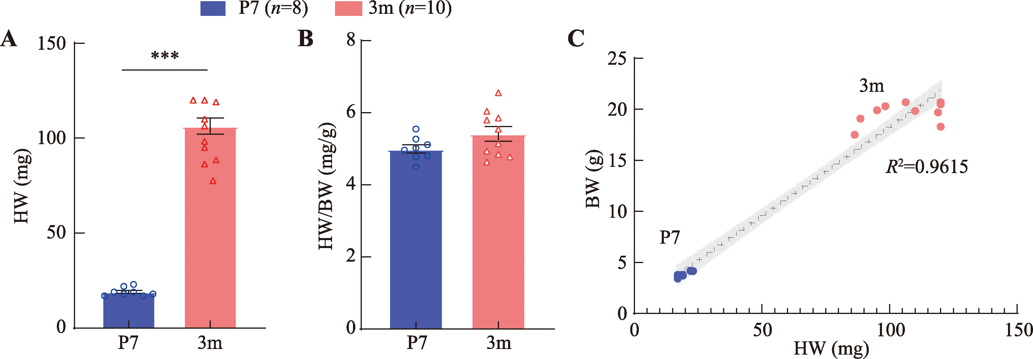
图2
小鼠心肌组织石蜡切片WGA染色分析 A:7日龄(P7)小鼠的心肌组织WGA染色结果;B:3月龄(3m)小鼠心肌组织WGA染色结果;C:P7、3m小鼠心肌细胞横截面积(μm²)的可视化分析。***P<0.001。"
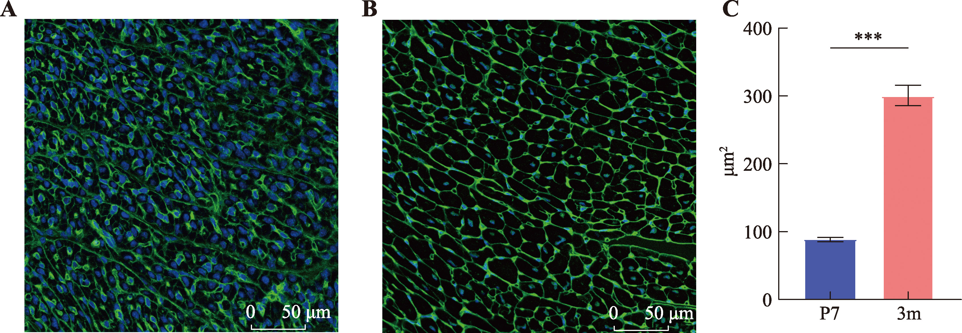
表2
转录组测序数据质量检测分析"
| 样本 | 原始读数 | 质控后读数 | 质控率 | Q30数据 |
|---|---|---|---|---|
| 9-2 | 74,869,328 | 71,669,772 | 95.72647961 | 94.88525 |
| 9-3 | 74,658,358 | 71,561,222 | 95.85158838 | 95.0681 |
| 9-5 | 78,257,522 | 74,637,762 | 95.37455326 | 94.8927 |
| 74 | 68,532,670 | 65,120,508 | 95.02111621 | 93.34445 |
| 48 | 79,189,892 | 76,434,652 | 96.52071757 | 94.78125 |
| C62 | 76,870,844 | 73,976,702 | 96.2350589 | 94.4907 |
| C63 | 81,374,030 | 76,251,440 | 93.70488349 | 94.839 |
表3
转录组测序基因组比对分析"
| 样本 | 比对率(%) |
|---|---|
| 9-2 | 93.09 |
| 9-3 | 93.56 |
| 9-5 | 93.44 |
| 74 | 90.83 |
| 48 | 90.08 |
| C62 | 93.27 |
| C63 | 92.78 |
图3
差异表达基因筛选和聚类分析 A:7日龄、3月龄小鼠心肌组织差异表达基因的火山图;B:7日龄、3月龄小鼠心肌组织差异表达基因的聚类热图。"
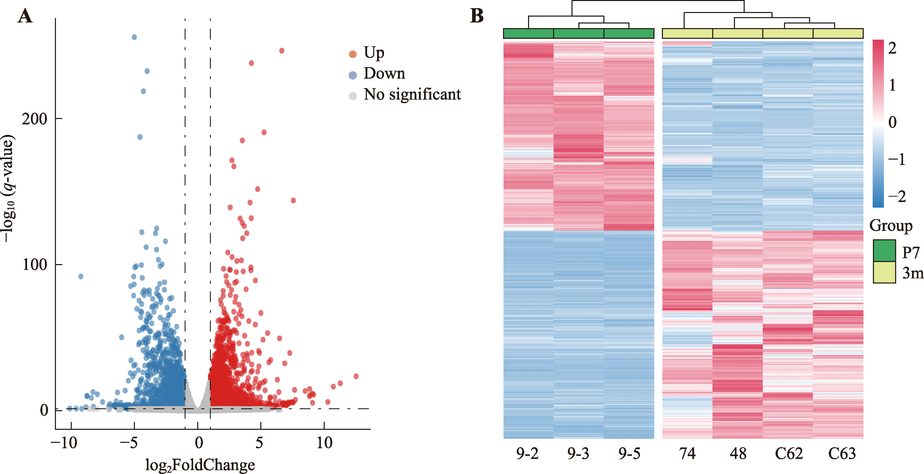
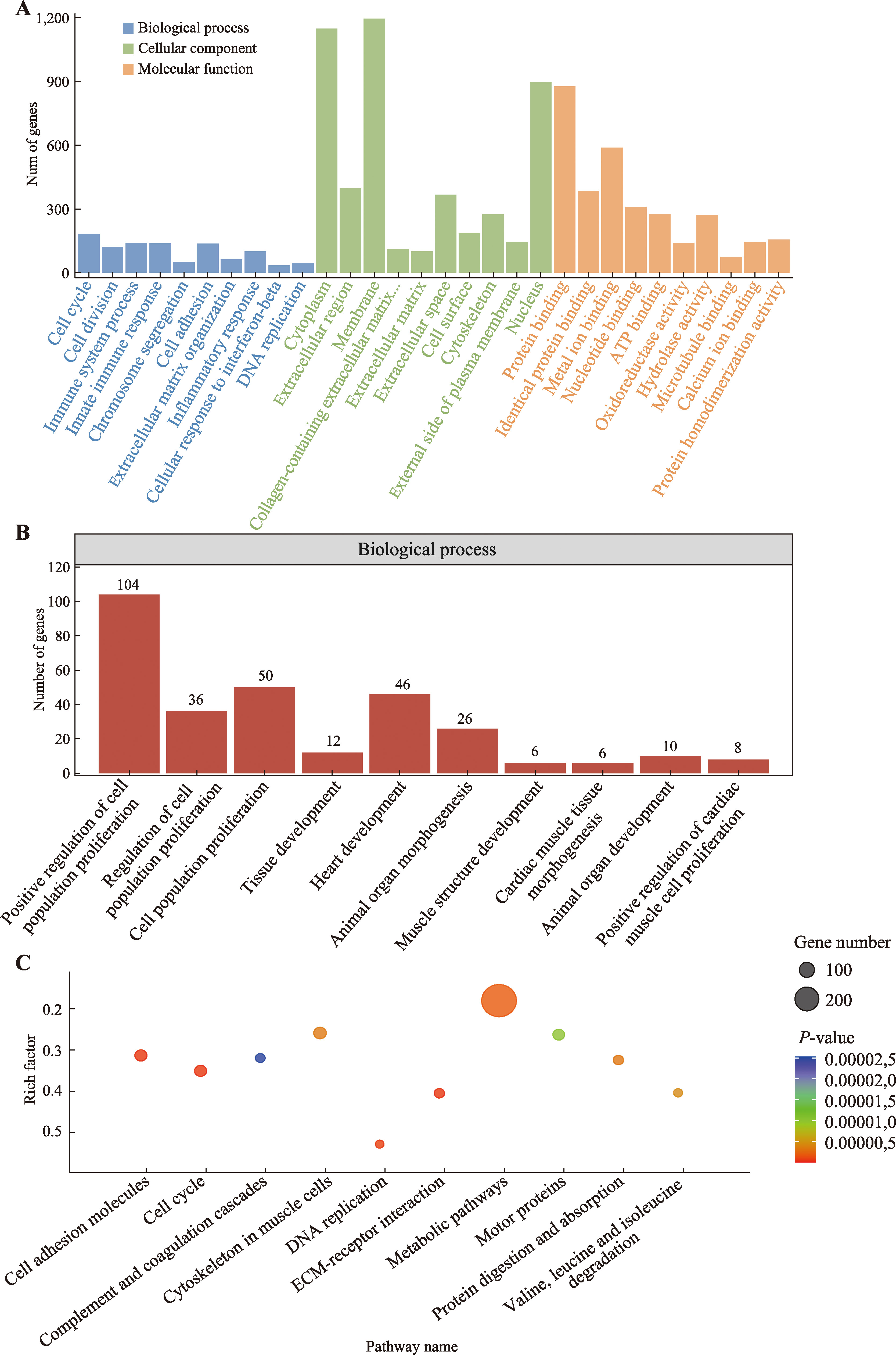
图4
差异表达基因的功能富集分析 A:差异表达基因的GO富集分析;B:与心脏发育、细胞增殖相关的GO条目;C:差异表达基因的KEGG通路富集分析。"
附表1
差异表达基因的GO分析"
| 类别 | GO编号 | 条目 | 数量 | P值 |
|---|---|---|---|---|
| BP | GO:0007049 | cell cycle | 182 | 4.06083E-37 |
| BP | GO:0051301 | cell division | 122 | 6.85875E-28 |
| BP | GO:0002376 | immune system process | 141 | 2.25781E-24 |
| BP | GO:0045087 | innate immune response | 138 | 9.96141E-24 |
| BP | GO:0007059 | chromosome segregation | 51 | 1.28172E-20 |
| BP | GO:0007155 | cell adhesion | 137 | 2.13134E-18 |
| BP | GO:0030198 | extracellular matrix organization | 62 | 1.79199E-17 |
| BP | GO:0006954 | inflammatory response | 100 | 3.63941E-16 |
| BP | GO:0035458 | cellular response to interferon-beta | 35 | 1.72508E-15 |
| BP | GO:0006260 | DNA replication | 44 | 7.87367E-15 |
| CC | GO:0005737 | cytoplasm | 1148 | 1.94244E-56 |
| CC | GO:0005576 | extracellular region | 397 | 1.46384E-45 |
| CC | GO:0016020 | membrane | 1195 | 2.89258E-32 |
| CC | GO:0062023 | collagen-containing extracellular matrix | 111 | 5.69624E-28 |
| CC | GO:0031012 | extracellular matrix | 100 | 5.86897E-27 |
| CC | GO:0005615 | extracellular space | 367 | 6.9155E-27 |
| CC | GO:0009986 | cell surface | 186 | 3.62299E-25 |
| CC | GO:0005856 | cytoskeleton | 275 | 6.68013E-25 |
| CC | GO:0009897 | external side of plasma membrane | 145 | 1.86746E-23 |
| CC | GO:0005634 | nucleus | 897 | 3.78546E-20 |
| MF | GO:0005515 | protein binding | 876 | 8.00395E-46 |
| MF | GO:0042802 | identical protein binding | 384 | 5.92756E-32 |
| MF | GO:0046872 | metal ion binding | 588 | 3.67755E-26 |
| MF | GO:0000166 | nucleotide binding | 310 | 1.38173E-22 |
| MF | GO:0005524 | ATP binding | 278 | 6.55811E-21 |
| MF | GO:0016491 | oxidoreductase activity | 141 | 2.05444E-20 |
| MF | GO:0016787 | hydrolase activity | 272 | 1.38026E-15 |
| MF | GO:0008017 | microtubule binding | 74 | 4.33152E-14 |
| MF | GO:0005509 | calcium ion binding | 144 | 6.90979E-14 |
| MF | GO:0042803 | protein homodimerization activity | 156 | 8.56528E-13 |
附表2
与心脏发育、器官发育、细胞增殖相关的GO条目"
| 编号 | 条目 | 数量 | P值 | 基因 |
|---|---|---|---|---|
| GO:0008284 | positive regulation of cell population proliferation | 104 | 1.22E-06 | Cdc20,Slc25a33,Hmgcr,Tff2,Crlf1,Has2,Cdk2,Efemp1,Ptn,Nanog,Folr2,Itgax,Tgfb2,Prc1,Rtkn2,Nrg4,Ets1,Tipin,Optn,Igf1,Sapcd2,Edn3,Agtr2,Cdk4,Sinhcaf,Acta2,Reg3g,Ccn2,Sfrp2,Bmp2Cntfr,Kif14,Dot1l,Cldn5,Id4,Pdgfa,Adrb2,Thrb,Flt3l,Fgf1,Aldh1a2,S100a1,Acer2,Lgals3,Cck,Ptgfr,Sox4,Mzb1,Prdx3,Ar,Sox9,Ptgs2,Mapk15,Pitx2,Il7r,Clec7a,Prlr,Csf1r,Akr1cl,Il6st,Apln,Cav3,Bst1,Epcam,Il15,Chp2,Cxcr3,Kit,Fn1,Epha4,Egf,Aqp11,Fgf7,Lamc2,Clu,Foxm1,Hif1a,Ctsh,Prkcz,Tnc,Ccdc117,Cdca7l,Cnot6,Osmr,Rad51b,Enpp2,Fgfr2,Csf1,Cx3cl1,Hmga2,Mlxipl,Cdc7,Igf2,Kif20b,Crh,Tnfsf13,Aif1,Ezh2,Marcksl1,Hipk2,Il6ra,Fbxo5,Hpse2,Fgf2 |
| GO:0042127 | regulation of cell population proliferation | 36 | 3.93E-04 | Ccdc88a,Ptgs2,Prg4,Osgin1,Dnmt1,Pf4,Pitx2,Sfrp2,Cxcl9,Junb,Gli3,Bcl6,Nr3c2,Sparc,Ptgs1,Serpine1,Tgfb2,Chek1,Emilin2,Cebpa,Emilin1,Sqle,Igf1,Nr5a2,Mzb1,Ezh2,Nfkbia,Clu,Bex4,Foxm1,Fas,Stat2,Txnip,Sox9,Hif1a,Cdca7 |
| GO:0008283 | immune effecell population proliferationctor process | 50 | 0.001055872 | Gdf11,Fam83d,Tff2,Csf1r,Prok1,Gli3,H3f3b,Rasgrp4,Kit,Glul,Ddr1,Egf,Igf1,Brca2,Hoxa3,Edn3,Foxm1,Mycn,Tacc2,Map7,Mcm7,St8sia1,Bmp7,Mcm10,Pax3,H19,Tlx1,Id4,Ptges,Pdgfa,Fgfr2,Bcl6,Aldh1a2,Tacc3,Hmga2,Il18,Cebpa,Cdkn2c,Flt3,Marcksl1,Hipk2,Atad5,Zbtb16,Sox9,Melk,Hpse2,Tbx20,Fgf2,Drd2,Mki67 |
| GO:0009888 | tissue development | 12 | 0.013249089 | Postn,Lamb1,Dhcr24,Lncpint,Lamc1,Lamc3,Lama4,Nr5a2,Gaa,Pax3,Ntng2,Lamc2 |
| GO:0007507 | heart development | 46 | 0.015057548 | Tgfb1i1,Odad2,Casp7,Pitx2,Col3a1,Bmp2,Ppara,Dnai1,Pax3,Robo1,Ptk7,Mb,Adamts6,Acan,Vcan,Myh10,Gli3,Mical2,Lox,Dnaaf3,Wt1,Frem2,Aldh1a2,Pdlim3,Gjc1,Adap2,Stra6,Hey2,Tgfb2,Mmp13,Tfdp2,Pde2a,Fn1,Sox4,Gja6,Pcna,Osr1,Fbn1,Sh3pxd2b,Aplnr,Prickle4,Sox9,Casp3,Col6a1,Mmp9,Adam19 |
| GO:0009887 | animal organ morphogenesis | 26 | 0.018261344 | Gdf11,Pitx2,Bmp2,Lamc3,Bmp7,Palb2,Asxl3,Pdgfa,Thrb,Fgfr2,Fgf1,Foxf2,Lama4,Ntng2,Gpc3,Wnt11,Igf2,Lamc1,Wnt5b,Fgf7,Hoxa3,Nkx2-1,Lamc2,Lamb1,Tcf21,Fgf2 |
| GO:0061061 | muscle structure development | 6 | 0.024757703 | Tgfb1i1,Prickle4,Ark2c,Col6a1,Hoxa2,Pdlim3 |
| cardiac muscle tissue morphogenesis | 6 | 0.024757703 | Gm1322,Angpt1,Bmp2,Egln1,Tbx20,Tcap | |
| GO:0048513 | animal organ development | 10 | 0.028652571 | Kit,Fmr1,Nrg2,Dand5,Flt1,Rai2,Pax3,Spry2,Csf1r,Tlx1 |
| GO:0060045 | positive regulation of cardiac muscle cell proliferation | 8 | 0.029630822 | Ccnb1,Fgfr2,Ncam1,Pim1,Fgf2,Tbx20,Hey2,Cdk1 |
附表3
上调差异表达基因显著富集的心肌发育相关通路"
| 编号 | 条目 | 数量 | P值 | 基因 |
|---|---|---|---|---|
| GO:0008016 | regulationofheartcontraction | 7 | 0.002745762 | Dmpk,Thrb,Kcnd2,S100a1,Hrc,Chrm2,Kcnd3 |
| GO:0060452 | positiveregulationofcardiacmusclecontraction | 4 | 0.014216607 | Adra1a,Ucn,Ace2,Ccn2 |
| mmu04260 | Cardiacmusclecontraction | 14 | 0.028156635 | Fxyd2,Atp1a2,COX3,Cacna2d1,Atp2a2,Cacna2d3,COX1,Cacna1s,CYTB,COX2,Hrc,Cox7a1,Atp2a1,Cox6a2 |
附表4
下调差异表达基因显著富集的心肌发育相关通路"
| 编号 | 条目 | 数量 | P值 | 基因 |
|---|---|---|---|---|
| GO:0007507 | heart development | 33 | 1.03E-04 | Odad2,Casp7,Pitx2,Col3a1,Bmp2,Ptk7,Robo1,Adamts6,Vcan,Acan,Myh10,Gli3,Mical2,Lox,Wt1,Frem2,Aldh1a2,Pdlim3,Gjc1,Hey2,Tgfb2,Fn1,Sox4,Pcna,Osr1,Fbn1,Sh3pxd2b,Aplnr,Sox9,Casp3,Col6a1,Tbx20,Adam19 |
| GO:0060045 | positive regulation of cardiac muscle cell proliferation | 7 | 0.002951524 | Ccnb1,Fgfr2,Ncam1,Pim1,Tbx20,Hey2,Cdk1 |
| GO:0055015 | ventricular cardiac muscle cell development | 4 | 0.010869872 | Myh10,Pitx2,Hey2,Cdk1 |
| GO:0010659 | cardiac muscle cell apoptotic process | 5 | 0.01482361 | Dynlt1f,Sfrp2,Dynlt1b,Hey2,Dynlt1a |
| GO:0060038 | cardiac muscle cell proliferation | 6 | 0.02682176 | Fgfr2,Myh10,Tenm4,2810429I04Rik,Hey2,Tgfb2 |
附表5
差异表达基因的KEGG通路富集分析"
| 编号 | 条目 | 数量 | P值 | 基因 |
|---|---|---|---|---|
| mmu04110 | Cell cycle | 55 | 1.10728E-10 | Cdc20,E2f1,Cdc25b,Bub1,Orc1,Mcm2,Cdk2,Mad2l1,Mtbp,Ccnb1,Plk1,Skp2,Tgfb2,Cdk1,Chek1,Ccne2,Esco2,Rbl1,Mcm6,Ndc80,Smc1b,Mcm4,Knl1,Gadd45a,Aurkb,E2f2,Cdc45,Pttg1,Cdk4,Ticrr,Ttk,Rbl2,Cdc6,Mcm7,Ccna1,Mcm3,Dbf4,Espl1,Ccna2,Cdc25c,Bub1b,Orc6,Tfdp2,Cdt1,Cdc7,Cdca5,Pcna,Cdkn2c,Ccne1,Mcm5,Sgo1,Fbxo5,Trip13,Pkmyt1,Ccnb2 |
| mmu04512 | ECM-receptor interaction | 36 | 3.9968E-09 | Thbs2,Lamc2,Chad,Lamb1,Col6a1,Comp,Col4a2,Col4a1,Itga2 |
| mmu04514 | Cell adhesion molecules | 56 | 8.23526E-09 | H2-Aa,Cldn3,H2-Ab1,Lrrc4b,H2-T24,Icam1,Mag,Itgb7,Selp,Sele,H2-Ob,Cldn5,Cd34,Cd274,Vcan,Tigit,Cd86,Nrxn3,Ctla4,Cd28,Sdc1,H2-Eb1,Cadm3,Madcam1,Slitrk4,Cldn15,H2-T23,H2-DMb1,Cldn11 |
| mmu03030 | DNA replication | 19 | 3.47378E-07 | Mcm7,Rfc3,Prim1,Pcna,Mcm3,Rfc5,Prim2,Rfc4,Lig1,Mcm6,Mcm2,Mcm4,Mcm5,Fen1,Pola2,Pole2,Rnaseh2b,Dna2,Pole |
| mmu01100 | Metabolic pathways | 296 | 1.56801E-06 | Dnmt1,Inpp5a,Lpin3,Hmgcr,Chsy3,Hddc2,St3gal5,Mtr,Cyp2e1,Adk,Selenbp1,Fmo1,Hsd11b1,Plcb2,Hpse,Bcat2,Gdpd3,Ptgs1,Cth,Phgdh,P4ha1,Echs1,Chpt1,Lalba,Nampt,Aldh4a1,Nat1,Ftcd,Acsl6,Ogdh,Hk2,Fmo2,E030003E18Rik,Cyp1a1,Nt5e,Abat,Hmgcs2,Oxct1,Ptgds,Bckdha,Sephs1,Tk1,Aldoc,Gaa,Dot1l,Pla2g12a,ND4L,Cpox,Scp2,Aoc1,Mpst,Aox3,St3gal6,Pomgnt2,Entpd2,Aldh1a2,Pdxk,Mocos,Nsd2,Aoc3,Papss1,Gda,Dck,Dctd,Nos1,Pfkm,Scd3,Tbxas1,Tpo,Scd2,Cyp26b1,Ehhadh,Extl1,Mccc2,Tha1,Me1,Ak9,Gsta13,Pfkfb1,Ptgs2,Hadhb,Ivd,Acss1,B4galt2,Scd4,COX3,Xdh,ATP6,Agpat2,Pdxp,Aldob,B3gnt5,Entpd5,COX1,Cyp26a1,Aldh6a1,COX2,Pla2g5,Ldhd,Ogdhl,P4ha3,Dgat2,Bst1,Gcdh,Acadsb,Pde2a,Glul,Pycr1,Auh,Lct,Pde6g,ND5,Gpld1,Alox5,St3gal3,Gstk1,Amd2,Gpam,Acot3,ND3,Hacd1,Tecr,Galnt15,Rrm1,Pnmt,Gpt2,Acsm5,Gsta1,Gstm1,St8sia1,Rimklb,Car5b,Mogat2,Fmo5,Alpl,Chac1,Ak4,Pde11a,Enpp2,Dpys,Gpt,Dhtkd1,Carnmt1,CYTB,Gsta3,Ethe1,Gstt2,Pld4,Gadl1,Khk,Pla2g2c,Cda,Cyp3a13,Lpin1,Cbs,Inmt,Gpx4,Hpse2,Nmnat3,Aspdh,Adcy9,Ddc,Gstt1,Mgat5b,Gstt3,Hk3,Gck,Nmrk2,Nqo1,L2hgdh,Xylt2,Suv39h1,Adi1,Cox6a2,Gsto2,Nnmt,Sqle,Gsto1,Pcx,Dbt,Kyat3,Cmbl,Asrgl1,Suox,Nmnat2,Tyms,Pipox,Pnpla2,Gnmt,Atp6v0e2,Dhfr,Cox6b2,Got1,Ahcyl2,Aldh1l1,Bckdhb,Amd1,Amy1,Psat1,Dnmt3b,Galnt16,Pafah1b3,Pnpla3,Hibadh,Rimkla,A4galt,Ptges,Dcxr,Dgkk,Pcca,Impa2,Gstm2,Gbgt1,Galnt5,Acer2,Gale,ND2,Elovl2,Mthfd1l,Sord,Aldh3b3,Inpp4b,Gsta2,Pde4c,Pank1,Acsl3,Pygb,Chsy1,Sds,Cyp2s1,Ggct,Ndst3,Maob,Hsd17b1,Nudt12,Acp5,Dhdh,Prodh,Galm,Chac2,Gpx3,Acot1,Pygm,Nt5c1a,Adh1,Mgst1,Kmt5c,P4ha2,Dglucy,Pcbd1,Ezh1,Chpf2,Aox1,Aspa,Mmut,Ckmt2,Acox1,Bdh1,Synj2,B3galt2,Pde7a,Aass,Mmab,Aldh2,Lrat,Dhcr24,Cds1,Mgat3,Cox6a1,Gstm7,Hnmt,Scd1,Acsbg3,Galk1,Pla2g7,Sqor,Rrm2,Upp1,Car15,Cyp27a1,Sardh,Pde4a,Gpx7,Suclg2,Ezh2,Adcy8,Pla2g2d,Ak7,Mgam,Ugt1a7c,Mccc1,Gstp2,Aldh1b1,Cox7a1,Gcat,ENSMUSG00000101904 |
| mmu04974 | Protein digestion and absorption | 35 | 3.16384E-06 | Col9a3,Eln,Col25a1,Col4a5,Col5a2,Col3a1,Col6a6,Col9a1,Col6a2,Col26a1,Slc36a2,Col9a2,Col1a1,Cpa2,Col27a1,Col8a1,Ace2,Col20a1,Col1a2,Col24a1,Prcp,Col12a1,Fxyd2,Col14a1,Atp1a2,Col18a1,Col11a1,Slc1a1,Col5a1,Col6a1,Col16a1,Kcne3,Col4a2,Cpa1,Col4a1 |
| mmu04820 | Cytoskeleton in muscle cells | 60 | 3.34597E-06 | Col3a1,Csrp2,Col9a2,Col1a1,Des,Sgca,Col27a1,Myh10,Tmod4,Sdc4,Pdlim3,Col1a2,Fn1,Myh8,Atp1a2,Fbn1,Fbln1,Col11a1,Thbs2,Col5a1,Myh14,Col6a1,Col4a2,Col4a1,Itgb7,Ankrd2,Thbs3,Col6a6,Col9a1,Diaph3,Csrp1,Col6a2,Myoz1,Itgb3,Vcan,Trim54,Col24a1,Lmod2,Sntb1,Myl9,Myot,Lmnb1,Gm1322,Actg2,Myoz2,Itgb6,Sdc1,Tcap,Fbn2,Sgcd,Tnni1,Comp,Nid2,Itga2 |
| mmu00280 | Valine, leucine and isoleucine degradation | 23 | 4.45307E-06 | Hadhb,Aldh2,Ivd,Bckdhb,Abat,Oxct1,Hmgcs2,Echs1,Auh,Bckdha,Dbt,Hibadh,Aox1,Mmut,Ehhadh,Pcca,Aldh6a1,Aox3,Mccc2,Mccc1,Bcat2,Aldh1b1,Acadsb |
| mmu04814 | Motor proteins | 52 | 1.04222E-05 | Kif15,Dnai2,Tuba4a,Myo7b,Myo1f,Dnai1,Dynlt1a,Kifc5b,Kif1c,Dnah12,Myh10,Myh8,Tuba1b,Tubb6,Dynlt1f,Dnah10,Myh14,Kif20a,Kif18b,Acta2,Dnah6,Tuba8,Kif11,Dnah9,Myo7a,Kifc1,Kif26a,Kif27,Kif18a,Dynlt1b,Kif14,Tubb4a,Tubb2b,Tubb5,Kif21a,Myo3b,Tube1,Kif23,Myl9,Kif7,Kif26b,Kif2c,Kif20b,Actg2,Cenpe,Kif24,Myo5b,Tnni1,Dnai3,Tuba1a,Kif4,Kif22 |
| mmu04610 | Complement and coagulation cascades | 30 | 2.53205E-05 | Bdkrb2,C1qb,C3,C6,Itgb2,Cfh,Vsig4,C4b,Serpina1b,Serpina1d,Cfd,Plat,Itgax,C1s1,Serpina1e,Serpine1,Itgam,C1qa,F7,C5ar1,C7,C1ra,Vwf,Serping1,C3ar1,Clu,Cd59b,F2rl3,F13a1,C1qc |
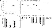
图5
候选基因的qRT-PCR验证 A:3个上调表达基因转录组测序差异倍数图;B:7个下调表达基因转录组测序差异倍数图;C:3个上调表达基因qRT-PCR变化倍数图;D:7个下调表达基因qRT-PCR变化倍数图。"

| [1] |
Mouse Genome Sequencing Consortium, Waterston RH, Lindblad-Toh K, Birney E, Rogers J, Abril JF, Agarwal P, Agarwala R, Ainscough R, Alexandersson M, An P, Antonarakis SE, Attwood J, Baertsch R, Bailey J, Barlow K, Beck S, Berry E, Birren B, Bloom T, Bork P, Botcherby M, Bray N, Brent MR, Brown DG, Brown SD, Bult C, Burton J, Butler J, Campbell RD, Carninci P, Cawley S, Chiaromonte F, Chinwalla AT, Church DM, Clamp M, Clee C, Collins FS, Cook LL, Copley RR, Coulson A, Couronne O, Cuff J, Curwen V, Cutts T, Daly M, David R, Davies J, Delehaunty KD, Deri J, Dermitzakis ET, Dewey C, Dickens NJ, Diekhans M, Dodge S, Dubchak I, Dunn DM, Eddy SR, Elnitski L, Emes RD, Eswara P, Eyras E, Felsenfeld A, Fewell GA, Flicek P, Foley K, Frankel WN, Fulton LA, Fulton RS, Furey TS, Gage D, Gibbs RA, Glusman G, Gnerre S, Goldman N, Goodstadt L, Grafham D, Graves TA, Green ED, Gregory S, Guigó R, Guyer M, Hardison RC, Haussler D, Hayashizaki Y, Hillier LW, Hinrichs A, Hlavina W, Holzer T, Hsu F, Hua A, Hubbard T, Hunt A, Jackson I, Jaffe DB, Johnson LS, Jones M, Jones TA, Joy A, Kamal M, Karlsson EK, Karolchik D, Kasprzyk A, Kawai J, Keibler E, Kells C, Kent WJ, Kirby A, Kolbe DL, Korf I, Kucherlapati RS, Kulbokas EJ, Kulp D, Landers T, Leger JP, Leonard S, Letunic I, Levine R, Li J, Li M, Lloyd C, Lucas S, Ma B, Maglott DR, Mardis ER, Matthews L, Mauceli E, Mayer JH, Mccarthy M, Mccombie WR, Mclaren S, Mclay K, Mcpherson JD, Meldrim J, Meredith B, Mesirov JP, Miller W, Miner TL, Mongin E, Montgomery KT, Morgan M, Mott R, Mullikin JC, Muzny DM, Nash WE, Nelson JO, Nhan MN, Nicol R, Ning ZM, Nusbaum C, O'Connor MJ, Okazaki Y, Oliver K, Overton-Larty E, Pachter L, Parra G, Pepin KH, Peterson J, Pevzner P, Plumb R, Pohl CS, Poliakov A, Ponce TC, Ponting CP, Potter S, Quail M, Reymond A, Roe BA, Roskin KM, Rubin EM, Rust AG, Santos R, Sapojnikov V, Schultz B, Schultz J, Schwartz MS, Schwartz S, Scott C, Seaman S, Searle S, Sharpe T, Sheridan A, Shownkeen R, Sims S, Singer JB, Slater G, Smit A, Smith DR, Spencer B, Stabenau A, Stange-Thomann N, Sugnet C, Suyama M, Tesler G, Thompson J, Torrents D, Trevaskis E, Tromp J, Ucla C, Ureta-Vidal A, Vinson JP, Von Niederhausern AC, Wade CM, Wall M, Weber RJ, Weiss RB, Wendl MC, West AP, Wetterstrand K, Wheeler R, Whelan S, Wierzbowski J, Willey D, Williams S, Wilson RK, Winter E, Worley KC, Wyman D, Yang S, Yang SP, Zdobnov EM, Zody MC, Lander ES. Initial sequencing and comparative analysis of the mouse genome. Nature, 2002, 420(6915): 520-562.
pmid: 12466850 |
| [2] |
Church DM, Goodstadt L, Hillier LW, Zody MC, Goldstein S, She XW, Bult CJ, Agarwala R, Cherry JL, Dicuccio M, Hlavina W, Kapustin Y, Meric P, Maglott D, Birtle Z, Marques AC, Graves T, Zhou SG, Teague B, Potamousis K, Churas C, Place M, Herschleb J, Runnheim R, Forrest D, Amos-Landgraf J, Schwartz DC, Cheng Z, Lindblad-Toh K, Eichler EE, Ponting CP, Mouse Genome Sequencing Consortium. Lineage-specific biology revealed by a finished genome assembly of the mouse. PLoS Biol, 2009, 7(5): e1000112.
pmid: 19468303 |
| [3] |
Gates H, Mallon AM, Brown SDM, EUMODIC Consortium. High-throughput mouse phenotyping. Methods, 2011, 53(4): 394-404.
pmid: 21185382 |
| [4] |
Peters LL, Robledo RF, Bult CJ, Churchill GA, Paigen BJ, Svenson KL. The mouse as a model for human biology: a resource guide for complex trait analysis. Nat Rev Genet, 2007, 8(1): 58-69.
pmid: 17173058 |
| [5] |
Mollova M, Bersell K, Walsh S, Savla J, Das LT, Park SY, Silberstein LE, Dos RC, Graham D, Colan S, Kühn B. Cardiomyocyte proliferation contributes to heart growth in young humans. Proc Natl Acad Sci USA, 2013, 110(4): 1446-1451.
pmid: 23302686 |
| [6] |
Leu M, Ehler E, Perriard JC. Characterisation of postnatal growth of the murine heart. Anat Embryol (Berl), 2001, 204(3): 217-224.
pmid: 11681801 |
| [7] |
Brendler J, Winter K, Lochhead P, Schulz A, Ricken AM. Histological differences between lumbar and tail intervertebral discs in mice. J Anat, 2022, 240(1): 84-93.
pmid: 34427936 |
| [8] |
Wong KC, Cao SS, Dong XL, Law MC, Chan TH, Wong MS. (-)-epiafzelechin protects against ovariectomy- induced bone loss in adult mice and modulate osteoblastic and osteoclastic ounctions in vitro. Nutrients, 2017, 9(5): 530.
pmid: 28531166 |
| [9] |
Jonas AM. The mouse in biomedical research. Physiologist, 1984, 27(5): 330-346.
pmid: 6393160 |
| [10] |
Wang ZN, Cui M, Shah AM, Ye WD, Tan W, Min YL, Botten GA, Shelton JM, Liu N, Bassel-Duby R, Olson EN. Mechanistic basis of neonatal heart regeneration revealed by transcriptome and histone modification profiling. Proc Natl Acad Sci USA, 2019, 116(37): 18455-18465.
pmid: 31451669 |
| [11] |
Bei YH, Chen C, Hua XJ, Yin MM, Meng XM, Huang ZZ, Qi WT, Su ZH, Liu C, Lehmann HI, Li GP, Huang Y, Xiao JJ. A modified apical resection model with high accuracy and reproducibility in neonatal mouse and rat hearts. NPJ Regen Med, 2023, 8(1): 9.
pmid: 36806296 |
| [12] |
Naqvi N, Li M, Calvert JW, Tejada T, Lambert JP, Wu JX, Kesteven SH, Holman SR, Matsuda T, Lovelock JD, Howard WW, Iismaa SE, Chan AY, Crawford BH, Wagner MB, Martin DI, Lefer DJ, Graham RM, Husain A. A proliferative burst during preadolescence establishes the final cardiomyocyte number. Cell, 2014, 157(4): 795-807.
pmid: 24813607 |
| [13] |
Alkass K, Panula J, Westman M, Wu TD, Guerquin-Kern JL, Bergmann O. No evidence for cardiomyocyte number expansion in preadolescent mice. Cell, 2015, 163(4): 1026-1036.
pmid: 26544945 |
| [14] |
Burrell JH, Boyn AM, Kumarasamy V, Hsieh A, Head SI, Lumbers ER. Growth and maturation of cardiac myocytes in fetal sheep in the second half of gestation. Anat Rec A Discov Mol Cell Evol Biol, 2003, 274(2): 952-961.
pmid: 12973719 |
| [15] |
Botting KJ, Wang KCW, Padhee M, Mcmillen IC, Summers-Pearce B, Rattanatray L, Cutri N, Posterino GS, Brooks DA, Morrison JL. Early origins of heart disease: low birth weight and determinants of cardiomyocyte endowment. Clin Exp Pharmacol Physiol, 2012, 39(9): 814-823.
pmid: 22126336 |
| [16] |
Rigaud VOCR, Hoy RC, Kurian J, Zarka C, Behanan M, Brosious I, Pennise J, Patel T, Wang T, Johnson J, Kraus LM, Mohsin S, Houser SR, Khan M. RNA-binding protein LIN28a regulates new myocyte formation in the heart through long noncoding RNA-H19. Circulation, 2023, 147(4): 324-337.
pmid: 36314132 |
| [17] |
Zhang HJ, Pei LJ, Ouyang ZH, Wang HC, Chen X, Jiang K, Huang SQ, Jiang R, Xiang YZ, Wei K. AP-1 activation mediates post-natal cardiomyocyte maturation. Cardiovasc Res, 2023, 119(2): 536-550.
pmid: 35640820 |
| [18] |
Eulalio A, Mano M, Dal Ferro M, Zentilin L, Sinagra G, Zacchigna S, Giacca M. Functional screening identifies miRNAs inducing cardiac regeneration. Nature, 2012, 492(7429): 376-381.
pmid: 23222520 |
| [19] |
Gan JY, Sonntag HJ, Tang MK, Cai DQ, Lee KKH. Integrative analysis of the developing postnatal mouse heart transcriptome. PLoS One, 2015, 10(7): e133288.
pmid: 26200114 |
| [20] |
Li F, Wang X, Capasso JM, Gerdes AM. Rapid transition of cardiac myocytes from hyperplasia to hypertrophy during postnatal development. J Mol Cell Cardiol, 1996, 28(8): 1737-1746.
pmid: 8877783 |
| [21] |
Lessard-Beaudoin M, Laroche M, Demers MJ, Grenier G, Graham RK. Characterization of age-associated changes in peripheral organ and brain region weights in C57BL/6 mice. Exp Gerontol, 2015, 63: 27-34.
pmid: 25597278 |
| [22] | Nirogi R, Goyal VK, Jana S, Pandey SK, Gothi A. What suits best for organ weight analysis: review of relationship between organ weight and body /brain weight for rodent toxicity studies. International Journal of Pharmaceutical Sciences and Research, 2014, 5(4): 1525-1532. |
| [23] |
Lopaschuk GD, Collins-Nakai RL, Itoi T. Developmental changes in energy substrate use by the heart. Cardiovasc Res, 1992, 26(12): 1172-1180.
pmid: 1288863 |
| [24] |
Bae J, Paltzer WG, Mahmoud AI. The role of metabolism in heart failure and regeneration. Front Cardiovasc Med, 2021, 8: 702920.
pmid: 34336958 |
| [25] |
Li X, Wu F, Günther S, Looso M, Kuenne C, Zhang T, Wiesnet M, Klatt S, Zukunft S, Fleming I, Poschet G, Wietelmann A, Atzberger A, Potente M, Yuan XJ, Braun T. Inhibition of fatty acid oxidation enables heart regeneration in adult mice. Nature, 2023, 622(7983): 619-626.
pmid: 37758950 |
| [26] |
Gao F, Liang T, Lu YW, Pu LB, Fu XY, Dong XX, Hong TT, Zhang F, Liu N, Zhou YX, Wang HK, Liang P, Guo YX, Yu H, Zhu W, Hu XY, Chen H, Zhou B, Pu WT, Mably JD, Wang JA, Wang DZ, Chen JH. Reduced mitochondrial protein translation promotes cardiomyocyte proliferation and heart regeneration. Circulation, 2023, 148(23): 1887-1906.
pmid: 37905452 |
| [27] |
Beura LK, Hamilton SE, Bi K, Schenkel JM, Odumade OA, Casey KA, Thompson EA, Fraser KA, Rosato PC, Filali-Mouhim A, Sekaly RP, Jenkins MK, Vezys V, Haining WN, Jameson SC, Masopust D. Normalizing the environment recapitulates adult human immune traits in laboratory mice. Nature, 2016, 532(7600): 512-516.
pmid: 27096360 |
| [28] |
Luan J, Xu H, Jin Z, Guan H, Gao XC, Gou XC, Xu LX. Analysis of the dynamic changes in the proportion of immune cells and the proportion of cells with stem cell characteristics in the corresponding immune cell population of C57 mice during the natural aging process. Immunol Res, 2021, 69(6): 520-532.
pmid: 34415527 |
| [29] |
Song XY, Zhang HH, Zhang YB, Goh B, Bao B, Mello SS, Sun XM, Zheng W, Gazzaniga FS, Wu M, Qu FF, Yin QZ, Gilmore MS, Oh SF, Kasper DL. Gut microbial fatty acid isomerization modulates intraepithelial T cells. Nature, 2023, 619(7971): 837-843.
pmid: 37380774 |
| [30] |
Wierstra I, Alves J. FOXM1, a typical proliferation- associated transcription factor. Biol Chem, 2007, 388(12): 1257-1274.
pmid: 18020943 |
| [31] |
Wierstra I. The transcription factor FOXM1 (Forkhead box M1): proliferation-specific expression, transcription factor function, target genes, mouse models, and normal biological roles. Adv Cancer Res, 2013, 118: 97-398.
pmid: 23768511 |
| [32] |
Kalin TV, Ustiyan V, Kalinichenko VV. Multiple faces of FoxM1 transcription factor: lessons from transgenic mouse models. Cell Cycle, 2011, 10(3): 396-405.
pmid: 21270518 |
| [33] |
Ye H, Kelly TF, Samadani U, Lim L, Rubio S, Overdier DG, Roebuck KA, Costa RH. Hepatocyte nuclear factor 3/fork head homolog 11 is expressed in proliferating epithelial and mesenchymal cells of embryonic and adult tissues. Mol Cell Biol, 1997, 17(3): 1626-1641.
pmid: 9032290 |
| [34] |
Bolte C, Zhang YF, Wang IC, Kalin TV, Molkentin JD, Kalinichenko VV. Expression of Foxm1 transcription factor in cardiomyocytes is required for myocardial development. PLoS One, 2011, 6(7): e22217.
pmid: 21779394 |
| [35] |
Ramakrishna S, Kim IM, Petrovic V, Malin D, Wang IC, Kalin TV, Meliton L, Zhao YY, Ackerson T, Qin YM, Malik AB, Costa RH, Kalinichenko VV. Myocardium defects and ventricular hypoplasia in mice homozygous null for the Forkhead Box M1 transcription factor. Dev Dyn, 2007, 236(4): 1000-1013.
pmid: 17366632 |
| [36] |
Zuppo DA, Missinato MA, Santana-Santos L, Li G, Benos PV, Tsang M. Foxm1 regulates cardiomyocyte proliferation in adult zebrafish after cardiac injury. Development, 2023, 150(6): dev201163.
pmid: 36846912 |
| [37] |
Sengupta A, Kalinichenko VV, Yutzey KE. FoxO1 and FoxM1 transcription factors have antagonistic functions in neonatal cardiomyocyte cell-cycle withdrawal and IGF1 gene regulation. Circ Res, 2013, 112(2): 267-277.
pmid: 23152492 |
| [38] |
Doi H, Iso T, Yamazaki M, Akiyama H, Kanai H, Sato H, Kawai-Kowase K, Tanaka T, Maeno T, Okamoto E, Arai M, Kedes L, Kurabayashi M. HERP1 inhibits myocardin- induced vascular smooth muscle cell differentiation by interfering with SRF binding to CArG box. Arterioscler Thromb Vasc Biol, 2005, 25(11): 2328-2334.
pmid: 16151017 |
| [39] |
Iso T, Sartorelli V, Chung G, Shichinohe T, Kedes L, Hamamori Y. HERP, a new primary target of Notch regulated by ligand binding. Mol Cell Biol, 2001, 21(17): 6071-6079.
pmid: 11486044 |
| [40] |
Donovan J, Kordylewska A, Jan YN, Utset MF. Tetralogy of fallot and other congenital heart defects in Hey2 mutant mice. Curr Biol, 2002, 12(18): 1605-1610.
pmid: 12372254 |
| [41] |
Koibuchi N, Chin MT. CHF1/Hey2 plays a pivotal role in left ventricular maturation through suppression of ectopic atrial gene expression. Circ Res, 2007, 100(6): 850-855.
pmid: 17332425 |
| [42] |
Ornitz DM, Itoh N. The fibroblast growth factor signaling pathway. Wiley Interdiscip Rev Dev Biol, 2015, 4(3): 215-266.
pmid: 25772309 |
| [43] |
Xie YL, Su N, Yang J, Tan QY, Huang S, Jin M, Ni ZH, Zhang B, Zhang DL, Luo FT, Chen HG, Sun XD, Feng JQ, Qi HB, Chen L. FGF/FGFR signaling in health and disease. Signal Transduct Target Ther, 2020, 5(1): 181.
pmid: 32879300 |
| [44] |
Matsuyama D, Kawahara K. Proliferation of neonatal cardiomyocytes by connexin43 knockdown via synergistic inactivation of p38 MAPK and increased expression of FGF1. Basic Res Cardiol, 2009, 104(6): 631-642.
pmid: 19377854 |
| [45] |
Jonker JW, Suh JM, Atkins AR, Ahmadian M, Li PP, Whyte J, He MX, Juguilon H, Yin YQ, Phillips CT, Yu RT, Olefsky JM, Henry RR, Downes M, Evans RM. A PPARgamma-FGF1 axis is required for adaptive adipose remodelling and metabolic homeostasis. Nature, 2012, 485(7398): 391-394.
pmid: 22522926 |
| [46] |
Madiai F, Hackshaw K. Expression of the mouse FGF-1 and FGF-1.A mRNAs during embryonic development and in the aging heart. Res Commun Mol Pathol Pharmacol, 2002, 112(1-4): 139-144.
pmid: 15080504 |
| [47] |
Novoyatleva T, Sajjad A, Pogoryelov D, Patra C, Schermuly RT, Engel FB. FGF1-mediated cardiomyocyte cell cycle reentry depends on the interaction of FGFR-1 and Fn14. FASEB J, 2014, 28(6): 2492-2503.
pmid: 24571920 |
| [48] |
Engel FB, Hsieh PCH, Lee RT, Keating MT. FGF1/p38 MAP kinase inhibitor therapy induces cardiomyocyte mitosis, reduces scarring, and rescues function after myocardial infarction. Proc Natl Acad Sci USA, 2006, 103(42): 15546-15551.
pmid: 17032753 |
| [49] |
Raju R, Palapetta SM, Sandhya VK, Sahu A, Alipoor A, Balakrishnan L, Advani J, George B, Kini KR, Geetha NP, Prakash HS, Prasad TS, Chang YJ, Chen LY, Pandey A, Gowda H. A network map of FGF-1/FGFR signaling system. J Signal Transduct, 2014, 2014: 962962.
pmid: 24829797 |
| [50] |
Cao HJ, Zhao LN, Yuan Y, Liao CY, Zeng WD, Li AY, Huang QF, Zhao YY, Fan YB, Jiang L, Song DD, Li S, Zhang B. Lipoamide attenuates hypertensive myocardial hypertrophy through PI3K/Akt-mediated Nrf2 signaling pathway. J Cardiovasc Transl Res, 2024, 17(4): 910-922.
pmid: 38334841 |
| [51] |
Qian WC, Yu DS, Zhang J, Hu QY, Tang CF, Liu PY, Ye P, Wang XL, Lv Q, Chen ML, Sheng L. Wogonin attenuates isoprenaline-induced myocardial hypertrophy in mice by suppressing the PI3K/Akt pathway. Front Pharmacol, 2018, 9: 896.
pmid: 30150938 |
| [52] |
Cao SM, Wang XY, Xing LJ, Zhang WG. Effects of long-term administration of bovine bone gelatin peptides on myocardial hypertrophy in spontaneously hypertensive rats. Nutrients, 2023, 15(24): 5021.
pmid: 38140281 |
| [53] |
Shao MJ, Zhuo CJ, Jiang RH, Chen GD, Shan JM, Ping J, Tian HJ, Wang LN, Lin CG, Hu LR. Protective effect of hydrogen sulphide against myocardial hypertrophy in mice. Oncotarget, 2017, 8(14): 22344-22352.
pmid: 28423592 |
| [54] |
Greulich F, Rudat C, Kispert A. Mechanisms of T-box gene function in the developing heart. Cardiovasc Res, 2011, 91(2): 212-222.
pmid: 21498422 |
| [55] |
Lu F, Langenbacher A, Chen JN. Tbx20 drives cardiac progenitor formation and cardiomyocyte proliferation in zebrafish. Dev Biol, 2017, 421(2): 139-148.
pmid: 27940156 |
| [56] |
Fang YB, Lai KS, She PL, Sun JJ, Tao WF, Zhong TP. Tbx20 induction promotes zebrafish heart regeneration by inducing cardiomyocyte dedifferentiation and endocardial expansion. Front Cell Dev Biol, 2020, 8: 738.
pmid: 32850848 |
| [57] |
Xiang FL, Guo MZ, Yutzey KE. Overexpression of tbx 20 in adult cardiomyocytes promotes proliferation and improves cardiac function after myocardial infarction. Circulation, 2016, 133(11): 1081-1092.
pmid: 26841808 |
| [58] |
Bersell K, Arab S, Haring B, Kuhn B. Neuregulin1/ErbB4 signaling induces cardiomyocyte proliferation and repair of heart injury. Cell, 2009, 138(2): 257-270.
pmid: 19632177 |
| [59] |
Lin ZQ, Zhou PZ, von Gise A, Gu F, Ma Q, Chen JH, Guo HD, van Gorp PRR, Wang DZ, Pu WT. Pi3kcb links Hippo-YAP and PI3K-AKT signaling pathways to promote cardiomyocyte proliferation and survival. Circ Res, 2015, 116(1): 35-45.
pmid: 25249570 |
| [60] |
Xin M, Kim Y, Sutherland LB, Murakami M, Qi XX, Mcanally J, Porrello ER, Mahmoud AI, Tan W, Shelton JM, Richardson JA, Sadek HA, Bassel-Duby R, Olson EN. Hippo pathway effector Yap promotes cardiac regeneration. Proc Natl Acad Sci USA, 2013, 110(34): 13839-13844.
pmid: 23918388 |
| [61] |
Heallen T, Morikawa Y, Leach J, Tao G, Willerson JT, Johnson RL, Martin JF. Hippo signaling impedes adult heart regeneration. Development, 2013, 140(23): 4683-4690.
pmid: 24255096 |
| [62] |
Sun LJ, Yu J, Qi S, Hao YW, Liu Y, Li ZW. Bone morphogenetic protein-10 induces cardiomyocyte proliferation and improves cardiac function after myocardial infarction. J Cell Biochem, 2014, 115(11): 1868-1876.
pmid: 24906204 |
| [63] |
Nipkow M, Wirthgen E, Luft P, Rebl A, Hoeflich A, Goldammer T. Characterization of igf1 and igf2 genes during maraena whitefish (Coregonus maraena) ontogeny and the effect of temperature on embryogenesis and igf expression. Growth Horm IGF Res, 2018, 40: 32-43.
pmid: 29723762 |
| [64] |
Jang JY, Song G, Pettit SM, Li QS, Song XS, Cai CL, Kaushal S, Li DQ. Epicardial hdac3 promotes myocardial growth through a novel microRNA pathway. Circ Res, 2022, 131(2): 151-164.
pmid: 35722872 |
| [65] |
Li P, Cavallero S, Gu Y, Chen THP, Hughes J, Hassan AB, Brüning JC, Pashmforoush M, Sucov HM. IGF signaling directs ventricular cardiomyocyte proliferation during embryonic heart development. Development, 2011, 138(9): 1795-1805.
pmid: 21429986 |
| [66] |
Pollak M. Insulin and insulin-like growth factor signalling in neoplasia. Nat Rev Cancer, 2008, 8(12): 915-928.
pmid: 19029956 |
| [67] |
Jung HJ, Suh Y. Regulation of IGF-1 signaling by microRNAs. Front Genet, 2014, 5: 472.
pmid: 25628647 |
| [68] |
Troncoso R, Ibarra C, Vicencio JM, Jaimovich E, Lavandero S. New insights into IGF-1 signaling in the heart. Trends Endocrinol Metab, 2014, 25(3): 128-137.
pmid: 38713091 |
| [69] |
Díaz Del Moral S, Benaouicha M, Muñoz-Chápuli R, Carmona R. The insulin-like growth factor signalling pathway in cardiac development and regeneration. Int J Mol Sci, 2021, 23(1): 234.
pmid: 35008660 |
| [70] |
Shoba L, An MR, Frank SJ, Lowe WJ Jr. Developmental regulation of insulin-like growth factor-I and growth hormone receptor gene expression. Mol Cell Endocrinol, 1999, 152(1-2): 125-136.
pmid: 10432230 |
| [71] |
Liu Q, Yan H, Dawes NJ, Mottino GA, Frank JS, Zhu H. Insulin-like growth factor II induces DNA synthesis in fetal ventricular myocytes in vitro. Circ Res, 1996, 79(4): 716-726.
pmid: 8831495 |
| [72] |
Hertig CM, Kubalak SW, Wang Y, Chien KR. Synergistic roles of neuregulin-1 and insulin-like growth factor-I in activation of the phosphatidylinositol 3-kinase pathway and cardiac chamber morphogenesis. J Biol Chem, 1999, 274(52): 37362-37369.
pmid: 10601306 |
| [73] |
Bhavsar PK, Brand NJ, Felkin LE, Luther PK, Cullen ME, Yacoub MH, Barton PJR. Clenbuterol induces cardiac myocyte hypertrophy via paracrine signalling and fibroblast-derived IGF-1. J Cardiovasc Transl Res, 2010, 3(6): 688-695.
pmid: 20577844 |
| [74] |
Wahyuni T, Kobayashi A, Tanaka S, Miyake Y, Yamamoto A, Bahtiar A, Mori S, Kametani Y, Tomimatsu M, Matsumoto K, Maeda M, Obana M, Fujio Y. Maresin-1 induces cardiomyocyte hypertrophy through IGF-1 paracrine pathway. Am J Physiol Cell Physiol, 2021, 321(1): C82-C93.
pmid: 34038245 |
| [75] |
Carè A, Catalucci D, Felicetti F, Bonci D, Addario A, Gallo P, Bang ML, Segnalini P, Gu YS, Dalton ND, Elia L, Latronico MVG, Høydal M, Autore C, Russo MA, Dorn GW 2nd, Ellingsen O, Ruiz-Lozano P, Peterson KL, Croce CM, Peschle C, Condorelli G. MicroRNA-133 controls cardiac hypertrophy. Nat Med, 2007, 13(5): 613-618.
pmid: 17468766 |
| [76] |
Younis S, Schönke M, Massart J, Hjortebjerg R, Sundström E, Gustafson U, Björnholm M, Krook A, Frystyk J, Zierath JR, Andersson L. The ZBED6-IGF2 axis has a major effect on growth of skeletal muscle and internal organs in placental mammals. Proc Natl Acad Sci USA, 2018, 115(9): E2048-E2057.
pmid: 29440408 |
| [1] | 朱前彬, 甘志承, 李晓翠, 张英杰, 赵合明, 黄先忠. 小鼠耳芥MAPKKK基因家族全基因组鉴定及进化与表达[J]. 遗传, 2022, 44(11): 1044-1055. |
| [2] | 李永光, 金玉环, 郭力, 艾昊, 李瑞宁, 黄先忠. 小鼠耳芥PEBP基因家族全基因组鉴定及表达分析[J]. 遗传, 2022, 44(1): 80-91. |
| [3] | 张为露, 冷奇颖, 郑嘉辉, AliHassanNawaz, 焦振海, 王府建, 张丽. 小鼠生长激素受体基因环状转录本的克隆及其表达规律[J]. 遗传, 2021, 43(9): 890-900. |
| [4] | 蒋婧, 赵安麒, 谢甜, 陈淑藯, 李劲松. 全基因组蛋白质的标签细胞和小鼠资源库建设[J]. 遗传, 2021, 43(7): 704-714. |
| [5] | 王剑飞, 韩俊海, 张子超. 孤独症谱系障碍小鼠模型行为学检测方法[J]. 遗传, 2021, 43(5): 501-519. |
| [6] | 朱文静, 刘志玮. MDMPR:小鼠发育代谢表型库[J]. 遗传, 2021, 43(4): 375-384. |
| [7] | 郝媛媛, 赵向前, 黄福灯, 李春寿. PPR蛋白在植物细胞器组分转录后调控中的作用机制[J]. 遗传, 2021, 43(11): 1050-1065. |
| [8] | 林珉婷, 赖璐璐, 赵淼, 林必玮, 姚香平. 利用CRISPR/Cas9 AAV系统构建纹状体Slc20a2基因敲除小鼠模型[J]. 遗传, 2020, 42(10): 1017-1027. |
| [9] | 于好强,孙福艾,冯文奇,路风中,李晚忱,付凤玲. 转录因子BES1/BZR1调控植物生长发育及抗逆性[J]. 遗传, 2019, 41(3): 206-214. |
| [10] | 刘钟颖,黄霞,李紫怡,杨子豪,袁白银. 肌动蛋白细胞骨架在小鼠第二生心区祖细胞部署发育中的作用[J]. 遗传, 2019, 41(2): 125-136. |
| [11] | 张高华, 于树涛, 王鹤, 王旭达. 高油酸花生发芽期低温胁迫转录组及差异表达基因分析[J]. 遗传, 2019, 41(11): 1050-1059. |
| [12] | 张豪, 张志鹏, 郭晓东, 马敏, 敖月, 刘旭, 马小燕, 梁浩, 郭旭东. cgVEGF164基因对小鼠毛囊生长的影响[J]. 遗传, 2019, 41(1): 76-84. |
| [13] | 孟晓伟, 汪洁, 马晴雯. 唐氏综合征小鼠模型的遗传背景和应用[J]. 遗传, 2018, 40(3): 207-217. |
| [14] | 谢晶, 范辰, 张景龙, 张仕强. Ash2l-1/Ash2l-2在小鼠胚胎干细胞中的表达特异性及互补效应[J]. 遗传, 2018, 40(3): 237-249. |
| [15] | 刘旭, 张平, 张晓枫, 李兴, 白宇, 贾克荣, 郭晓东, 张豪, 马晓燕, 仓明, 刘东军, 郭旭东. 利用CRISPR/Cas9系统构建FGF21基因敲除小鼠模型[J]. 遗传, 2018, 40(1): 66-74. |
| 阅读次数 | ||||||
|
全文 |
|
|||||
|
摘要 |
|
|||||

www.chinagene.cn
备案号:京ICP备09063187号-4
总访问:,今日访问:,当前在线:
